A team led by engineers at the University of California, San Diego has 3D-printed a tissue that closely mimics the human liver’s sophisticated structure and function. The new model could be used for patient-specific drug screening and disease modeling. The work was published the week of Feb. 8 in the online early edition of Proceedings of the National Academy of Sciences.
Researchers said the advance could help pharmaceutical companies save time and money when developing new drugs.
“It typically takes about 12 years and $1.8 billion to produce one FDA-approved drug,” said Shaochen Chen, NanoEngineering professor at the UC San Diego Jacobs School of Engineering. “That’s because over 90 percent of drugs don’t pass animal tests or human clinical trials. We’ve made a tool that pharmaceutical companies could use to do pilot studies on their new drugs, and they won’t have to wait until animal or human trials to test a drug’s safety and efficacy on patients. This would let them focus on the most promising drug candidates earlier on in the process.”
Chen and Shu Chien, a professor of Medicine and Bioengineering, director of the Institute of Engineering in Medicine at UC San Diego and recipient of a National Medal of Science, are co-senior authors of the study.
The liver plays a critical role in how the body metabolizes drugs and produces key proteins. This is why liver models are increasingly being developed in the lab as platforms for drug screening. However, existing models so far lack both the complex micro-architecture and diverse cell makeup of a real liver.
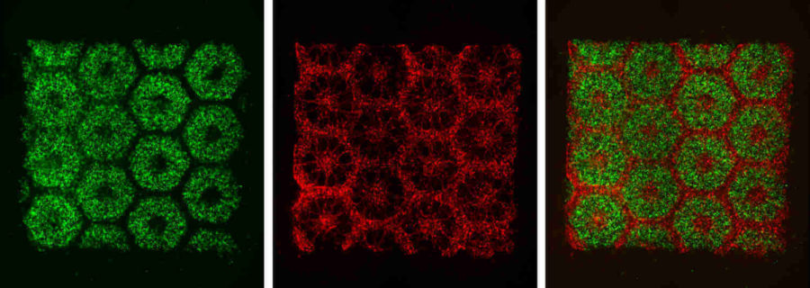
The UC San Diego team engineered a human liver tissue model that more closely resembles the real thing—a diverse combination of liver cells and supporting cells systematically organized in a hexagonal pattern.
“We’ve engineered a functioning liver tissue that matches what you’d see under a microscope,” said Chen.
“The liver is unique in that it receives a dual blood supply with different pressures and chemical constituents. Our model has the potential of reproducing this intricate blood supply system, thus providing unprecedented understanding of the complex coupling between circulation and metabolic functions of the liver in health and disease,” said Chien, who studies how blood flow and pressure affect blood vessels.
To do this, the team employed a novel bioprinting technology developed in Chen’s lab, which can rapidly produce complex 3D microstructures that mimic the sophisticated features found in biological tissues. The liver tissue was printed in two steps. First, the team printed a honeycomb pattern of 900-micrometer-sized hexagons, each containing liver cells derived from human induced pluripotent stem cells. An advantage of human induced pluripotent stem cells is they are patient-specific, which makes them ideal materials for building patient-specific drug screening platforms. And since these cells are derived from a patient’s skin cells, researchers don’t need to extract any cells from the liver to build liver tissue.
In the next step, endothelial and mesenchymal supporting cells were printed in the spaces between the stem-cell-containing hexagons.
The entire structure—a 3 × 3 millimeter square, 200 micrometers thick—takes just seconds to print. This is a vast improvement over other methods to print liver models, which typically take hours.
The structure was cultured in vitro for at least 20 days. The researchers then tested the resulting tissue’s ability to perform various liver functions, such as albumin secretion and urea production, and compared it to other models. They found that their model was able to maintain these functions over a longer time period than other liver models. Their model also expressed a relatively higher level of a key enzyme that’s considered to be involved in metabolizing many of the drugs administered to patients.
“I think that this will serve as a great drug screening tool for pharmaceutical companies and that our 3D bioprinting technology opens the door for patient-specific organ printing in the future,” said Chen.
“The liver tissue constructed by this novel 3D printing technology will also be extremely useful in reproducing in vitro disease models such as hepatitis, cirrhosis, and cancer,” added Chien. “Such realistic models will be invaluable for the study of the pathophysiology and metabolic abnormalities in these diseases and the efficacy of drug therapies.”