Researchers of the Max Planck Institute of Molecular Cell Biology and Genetics in Dresden report how they managed to capture detailed three-dimensional images of cardiac dynamics in zebrafish. The novel approach: They combine high-speed Selective Plane Illumination Microscopy (SPIM) and clever image processing to reconstruct multi-view movie stacks of the beating heart. Furthermore, they have developed a method of generating high-resolution static reconstructions of the zebrafish’s heart: the Dresden research team used optogenetics to stop the beating heart by illuminating it with light.
Non-periodic phenomena such as irregularly beating hearts and the flow of blood cells are resolved by high-speed volume scanning using a liquid lens. This work is set to be key in our understanding of congenital heart defects as well in future experiments on cardiac function and development. Until recently, available microscopes were too slow to capture a beating heart in 3D. Now, the team led by research group leader Jan Huisken at the Max Planck Institute of Molecular Cell Biology and Genetics has developed a high-speed, selective plane illumination microscope that manages to do just that. By gently illuminating the fish heart with a thin light sheet and observing the emitted fluorescence with a fast and sensitive camera the researchers have achieved fast, non-invasive imaging of labelled heart tissue. The process involves taking multiple movies, each covering individual planes of the heart (movie stacks), then using the correlations between the individual planes to generate a synchronised, dynamic 3D image of the beating heart.
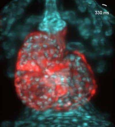
The team also obtained static high-resolution reconstructions by briefly stopping the heart with optogenetics. This procedure does not harm the fish – zebrafish embryos can survive a cardiac arrest of several hours. “These renderings allow us to further follow characteristic structures of the heart throughout the cardiac cycle,” says Michaela Mickoleit, PhD student who performed the experiments in Huisken’s lab. For instance, they now can clearly observe cardiac contractions or the distance between endo- and myocardium throughout the heartbeat.
By manipulating the exposure time and magnification of the images, better resolution could be achieved and fine details such as sarcomeres and filamentous actin could also be resolved. Finally, they then also went on to resolve non-periodic phenomena by high-speed volume scanning with a liquid lens. For the first time, it has become possible to also image diseased hearts that exhibit arrhythmia – exciting news for cardiologists.
The team at the Max Planck Institute of Molecular Cell Biology and Genetics has developed a fantastic array of tools to image the heart in vivo, ranging from static to ultra-high-speed images. Their work offers potentially revolutionary insights into the cellular structure of the beating heart and are set to further improve our knowledge of congenital heart defects.