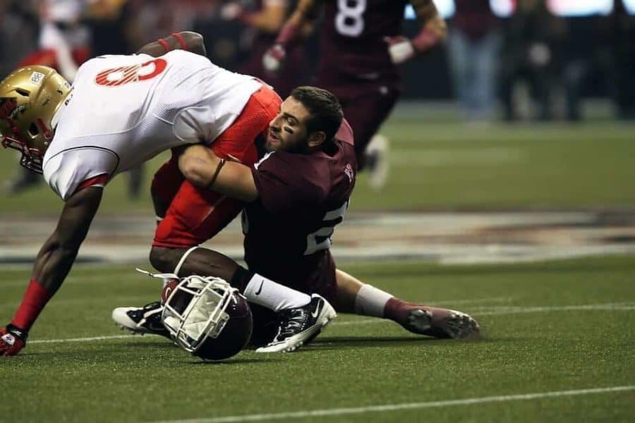
Mild head trauma in adolescents and adults who participate in contact sports damages the barrier that protects the brain from pathogens and toxins, according to researchers at the Stanford University School of Medicine and Trinity College in Dublin.
If the results of their small pilot study hold up, the brain imaging technique employed by the researchers could be used to monitor athletes who’ve taken a blow to the head and determine when they’re safe to resume contact sports.
In the study, which was published online Sept. 5 in the Journal of Neurotrauma, scientists scanned the brains of adolescent and adult rugby players with a special type of magnetic resonance imaging. They found damage to the protective barrier that separates the brain from bloodborne pathogens and toxins in roughly half of adolescent rugby players after a full season — even those who did not report a concussion. Professional mixed martial arts fighters showed similar damage after a fight.
The link between repeated head trauma and neurodegenerative disease continues to fuel public interest and debate. And though the new study draws no definitive conclusions, it shows that mild blows to the head may at least temporarily weaken an important brain barrier.
“This is some of the first evidence, certainly in kids, to show disruption of the blood-brain barrier even in the absence of concussion,” said David Camarillo, PhD, assistant professor of bioengineering at Stanford and co-senior author of the study.
Barrier breakdown
Most traumatic brain injuries are mild. That may sound like an oxymoron, but mild traumatic brain injuries only temporarily affect normal brain function.
The current diagnosis for mild head trauma is based on a temporary change in awareness and responsiveness, short-term amnesia, headache or general difficulty thinking clearly. But it can be difficult to reliably spot these symptoms during contact sports.
Researchers at Stanford and Trinity College teamed up to find a more objective way to pinpoint mild head trauma. The Dublin contingent had previously studied brains from patients afflicted with chronic traumatic encephalopathy, a neurodegenerative disease. Brains from these patients had a defunct blood-brain barrier — a border that allows oxygen and nutrients to pass into the brain while blocking larger molecules. This barrier operates a lot like a teabag, which lets water through but holds leaves in place.
The researchers wondered if mild head trauma damaged the blood-brain barrier. So they recruited a group of rugby players and scanned their brains with magnetic resonance imaging, or MRI, after a season or single match. To see if the blood-brain barrier was intact, Dublin researchers injected participants with a commonly used MRI contrast agent. Little to no contrast agent will show up in the brain of a person with an intact barrier.
Ten of 19 adolescent rugby players showed signs of blood-brain barrier disruption by the end of the season. The barrier breakdown appeared on the scans as red blips concentrated throughout the inner regions of the brain. The researchers also scanned eight college rugby players after a match and saw disruptions of the barrier in two of them. Notably, the injuries most study participants experienced were below the current bar for mild head trauma since they did not suffer a concussion.
“We’ve always been interested in studying rugby,” said co-author Gerald Grant, MD, professor of neurosurgery and a concussion expert at Stanford’s Children’s Health, whose lab studies the blood-brain barrier. “People have this sense that rugby must be safer because you’re usually avoiding head contact much more than in football since you’re not as protected.”
A productive partnership
Based on these initial findings, the researchers hypothesized that impact forces were damaging the blood-brain barrier. But they needed a way to precisely measure those forces. And they wanted to test other contact sports, as well.
Enter Camarillo, whose lab has developed a mouth guard that tracks speed, acceleration and force at nearly 10,000 measurements per second. That level of speed and precision was vital for the next contact sport the researchers studied: mixed martial arts. The scientists recruited five professional mixed martial arts fighters, who wore the mouth guards during fights and had their brains scanned before and after fights.
Post-fight MRI scans showed increased blood-brain barrier breakdown, just as the researchers observed in rugby players. And the researchers found that certain measurements from the mouth guards correlated with the level of blood-brain barrier disruption seen by MRI.
“This suggests there might be some combination of number of blows and severity of blows that might explain blood-brain barrier injury,” Camarillo said.
Not every hit that looked bad — such as when a fighter was knocked out during the first two minutes — damaged the blood brain barrier, emphasizing the value of the mouth guard measurements to record the forces of injury.
The findings, Camarillo said, are further evidence that the longstanding model of concussion is likely too simplistic. The current paradigm is that the brain slams into the skull, recoils and slams again on the opposite side. According to this model, most brain damage should occur along the outer surface of the brain. But recent evidence — including the present study — suggests that head trauma’s effects are felt much deeper in the brain.
Going forward, the researchers plan to conduct a similar study in a larger cohort. They’re also interested in determining whether the blood-brain barrier disruptions they observed heal on their own and, if so, how long that takes.
While the findings are still at an early stage, the imaging technique used in this study — perhaps in conjunction with blood biomarkers — could one day be used to figure out how much damage an athlete has sustained, and when he or she can return to play, according to Grant.
“I think we’re ignoring many of these kids who are experiencing these injuries throughout the season but aren’t aware of them, or they have no symptoms,” Grant said. “Maybe this study can help us figure out how to better classify some of these kids and figure out if they are truly safe to go back and play.”
Camarillo and Grant are members of Stanford Bio-X, the Stanford Maternal & Child Health Research Institute and the Wu Tsai Neurosciences Institute at Stanford.
Other Stanford co-authors of the study are postdoctoral scholar Yuzhe Liu, PhD, and former postdoctoral scholar Chiara Giordano, PhD.
Researchers at the Ben-Gurion University of the Negev in Israel also contributed to the study.
This study was supported by Science Foundation Ireland, The St. James’s Hospital Foundation, the National Institute of Neurological Disorders and Stroke and the Ellen Mayston Bates bequest to the Trinity Foundation.
Stanford’s Department of Bioengineering also supported the work.


