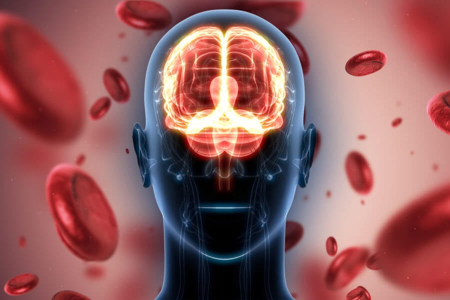
While neurons and glial cells are by far the most numerous cells in the brain, many other types of cells play important roles. Among those are cerebrovascular cells, which form the blood vessels that deliver oxygen and other nutrients to the brain.
Those cells, which comprise only 0.3 percent of the brain’s cells, also make up the blood-brain barrier, a critical interface that prevents pathogens and toxins from entering the brain, while allowing critical nutrients and signals through. Researchers from MIT have now performed an extensive analysis of these difficult-to-find cells in human brain tissue, creating a comprehensive atlas of cerebrovascular cell types and their functions.
Their study also revealed differences between cerebrovascular cells from healthy people and people suffering from Huntington’s disease, which could offer new targets for potential ways to treat Huntington’s disease. Breakdown of the blood-brain barrier is associated with Huntington’s and many other neurodegenerative diseases, and often occurs years before any other symptoms appear.
“We think this might be a very promising route because the cerebrovasculature is much more accessible for therapeutics than the cells that lie inside the blood-brain barrier of the brain,” says Myriam Heiman, an associate professor in MIT’s Department of Brain and Cognitive Sciences and a member of the Picower Institute for Learning and Memory.
Heiman and Manolis Kellis, a professor of computer science in MIT’s Computer Science and Artificial Intelligence Laboratory (CSAIL) and a member of the Broad Institute of MIT and Harvard, are the senior authors of the study, which appears today in Nature. MIT graduate students Francisco Garcia in the Department of Brain and Cognitive Sciences, and Na Sun in the Department of Electrical Engineering and Computer Science, are the lead authors of the paper.
A comprehensive atlas
Cerebrovascular cells make up the network of blood vessels that deliver oxygen and nutrients to the brain, and they also help to clear out debris and metabolites. Dysfunction of this irrigation system is believed to contribute to the buildup of harmful effects seen in Huntington’s disease, Alzheimer’s, and other neurodegenerative diseases.
Many types of cells are found in the cerebrovasculature, but because they make up such a small fraction of the cells in the brain, it has been difficult to obtain enough cells to perform large-scale analyses with single-cell RNA sequencing. This kind of study, which allows the gene expression patterns of individual cells to be deciphered, offers a great deal of information on the functions of specific cell types, based on which genes are turned on in those cells.
For this study, the MIT team was able to obtain over 100 human postmortem brain tissue samples, and 17 healthy brain tissue samples removed during surgery performed to treat epileptic seizures. That brain surgery tissue came from younger patients than the postmortem samples, enabling the researchers to also recognize age-associated differences in the vasculature. The researchers enriched the brain surgery samples for cerebrovascular cells using centrifugation, and ran postmortem sample cells through a computational “sorting” pipeline that identified cerebrovascular cells based on certain markers that they express.
The researchers performed single-cell RNA-sequencing on more than 16,000 cerebrovascular cells, and used the cells’ gene-expression patterns to classify them into 11 different subtypes. These types included endothelial cells, which line the blood vessels; mural cells, which include pericytes, found in the walls of capillaries, and smooth muscle cells, which help regulate blood pressure and flow; and fibroblasts, a type of structural cell.
“This study allowed us to zoom in to this incredibly central cell type that facilitates all of the functioning of the brain,” Kellis says. “What we’ve done here is understand these building blocks and this diversity of cell types that make up the vasculature in unprecedented resolution, across hundreds of individuals.”
The researchers also found evidence for a phenomenon known as zonation. This means that the endothelial cells that line the blood vessels express different genes depending on where they are located — in an arteriole, capillary, or venule. Furthermore, among the hundreds of genes they identified that are expressed differently in the three zones, only about 10 percent of them are the same as the zonated genes that have been previously seen in the mouse cerebrovasculature.
“We saw a lot of human specificity,” Heiman says. “What our study provides is a list of markers and insights into gene function in these three different regions. These are things that we believe are important to see from a human cerebrovasculature perspective, because the conservation between species is not perfect.”
Barrier breakdown
The researchers also used their new vasculature atlas to analyze a set of postmortem brain tissue samples from disease patients, demonstrating its broad usefulness. They focused on Huntington’s disease, where cerebrovasculature abnormalities include leakiness of the blood-brain barrier and a higher density of blood vessels. These symptoms usually appear before any of the other symptoms associated with Huntington’s, and can be seen using functional magnetic resonance imaging (fMRI).
In this study, the researchers found that cells from Huntington’s patients showed many changes in gene expression compared to healthy cells, including a decrease in the expression of the gene for MFSD2A, a key transporter that restricts the passage of lipids across the blood-brain barrier. They believe that the loss of that transporter, along with other changes they observed, could contribute to increased leakiness of the barrier.
They also found upregulation of genes involved in the Wnt signaling pathway, which promotes new blood vessel growth and that endothelial cells of the blood vessels showed unexpectedly strong immune activation, which may further contribute to blood-brain barrier dysregulation.
Because cerebrovascular cells can be accessed through the bloodstream, they could make an enticing target for possible treatments for Huntington’s and other neurodegenerative diseases, Heiman says. The researchers now plan to test whether they might be able to deliver potential drugs or gene therapy to these cells, and study what therapeutic effect they might have, in mouse models of Huntington’s disease.
“Given that cerebrovascular dysfunction arises years before more disease-specific symptoms, perhaps it’s an enabling factor for disease progression,” Heiman says. “If that’s true, and we can prevent that, that could be an important therapeutic opportunity.”
The researchers also plan to analyze more of the RNA-sequencing data from their tissue samples, beyond the cerebrovascular cells that they examined in this paper.
“Our goal is to build a systematic single-cell map to navigate brain function in health, disease, and aging across thousands of human brain samples,” Kellis says. “This study is one of the first bite-sized pieces of this atlas, looking at 0.3 percent of cells. We are actively analyzing the other 99 percent in multiple exciting collaborations, and many insights continue to lie ahead.”
The research was funded by the Intellectual and Developmental Disability Research Center and Rosamund Stone Zander Translational Neuroscience Center at Boston Children’s Hospital, a Picower Institute Innovation Fund Award, a Walter B. Brewer MIT Fund Award, the National Institutes of Health, and the Cure Alzheimer’s Fund.

