“Seeing the unseen” may sound like a science fiction movie theme, but it’s actually the real-life mission of USDA scientists who use special high-powered microscopes to view microscopic organisms that play a big role in agriculture.
The facility where these scientists produce the images of the unseen world–from fungal spores to plant cells–is called the Electron and Confocal Microscopy Unit (ECMU) and it’s operated in Beltsville, Md., by USDA’s Agricultural Research Service (ARS).
Researchers often request the images to aid them in their studies, and to illustrate papers, posters and grant proposals. Outside research organizations have also sought out the expertise of ECMU director Gary Bauchan and his colleagues in producing the images, as well as the skill with which they ensure the integrity of specimens and samples submitted to them.
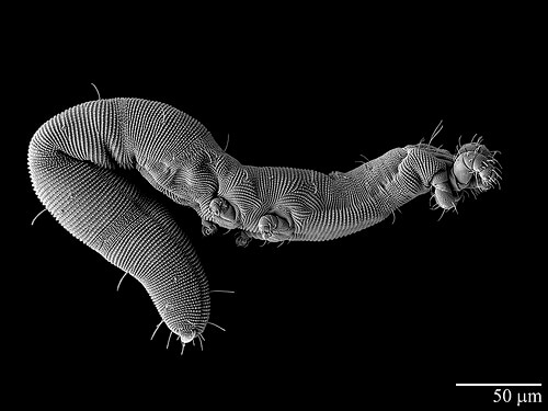
In December 2013, for example, Ohio State University (OSU) doctoral student Samuel Bolton finished a 12-month assignment working with Bauchan and ARS entomologist Ron Ochoa to identify and describe a new, worm-like species of mite belonging to the family Nematalycidae.
The mite, dubbed Osperalycus tenerphagus, “is the first species of this weird family to be described in over 40 years,” said Bolton, who studies mite systematics under the guidance of OSU professor Hans Klompen in Columbus, Ohio.
Bolton literally dug up the species while excavating loam soil across the street from OSU’s Museum of Biological Diversity. This was unusual because mites in the Nematalycidae family are mostly found in sandy habitats (dunes, beaches, desert soils).
Because the mites were worm-like and very soft-bodied, the only method for viewing the creatures in their natural state was to freeze them and observe them frozen. Thus, before imaging, the mites were placed on small, copper plates and then plunged into liquid nitrogen at minus 320 degrees Fahrenheit. Next, the specimens were thinly coated in platinum and positioned on a cold stage at minus 202 degrees Fahrenheit within a low-temperature scanning electron microscope. There, they were imaged at magnifications of up to 30,000 times.
“We are confident we have by far and away the best images that have ever been captured of this strange looking mite,” says Bolton. He, Bauchan, Ochoa and Klompen have published their discovery in The Journal of Natural History.