Harvard neuroscientists have made a discovery that turns 160 years of neuroanatomy on its head.
Myelin, the electrical insulating material long known to be essential for the fast transmission of impulses along the axons of nerve cells, is not as ubiquitous as thought, according to a new work lead by Professor Paola Arlotta of the Harvard Stem Cell Institute (HSCI) and the University’s Department of Stem Cell and Regenerative Biology, in collaboration with Professor Jeff Lichtman, of Harvard’s Department of Molecular and Cellular Biology.
“Myelin is a relatively recent invention during evolution,” says Arlotta. “It’s thought that myelin allowed the brain to communicate really fast to the far reaches of the body, and that it has endowed the brain with the capacity to compute higher level functions.” In fact, loss of myelin is a feature of a number of devastating diseases, including multiple sclerosis and schizophrenia.
But the new research shows that despite myelin essential roles in the brain, “some of the most evolved, most complex neurons of the nervous system have less myelin than older, more ancestral ones” Arlotta, co-director of the HSCI neuroscience program, says.
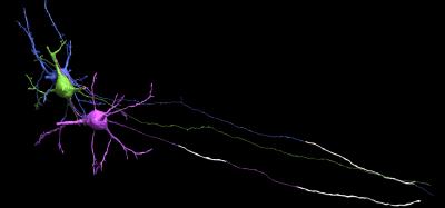
What this means, Arlotta says, is that the higher in the cerebral cortex one looks – the closer to the top of the brain, which is its most evolved region – the less myelin one finds. Not only that, but “neurons in this part of the brain display a brand new way of positioning myelin along their axons that has not been previously seen. They have ‘intermittent myelin’ with long axon tracts that lack myelin interspersed among myelin-rich segments.
Arlotta continues: “contrary to the common assumptions that neurons use a universal profile of myelin distribution on their axons, the work indicate that different neurons choose to myelinate their axons differently. In classic neurobiology textbooks myelin is represented on axons as a sequence of myelinated segments separated by very short nodes that lack myelin. This distribution of myelin was tacitly assumed to be always the same, on every neuron, from the beginning to the end of the axon. This new work finds this not to be the case.”
The results of the research by Arlotta and post doctoral fellow Giulio Srubek Tomassy, the first author on the report, are published in the latest edition of Science, the journal of the American Association for the Advancement of Science.
The paper is accompanied by a “Perspective” by R. Douglas Fields, of the Eunice Kennedy Shriver National Institute of Child Health and Human Development, at the National Institutes of Health, who says that Arlotta and Tomassy’s findings raise important questions about the purpose of myelin, “are likely to spark new concepts about how information is transmitted and integrated in the brain.”
Arlotta and Tomassy collaborated closely on the new work with postdoctoral fellow Daniel Berger of the Lichtman group, which generated one of the two massive electron microscopy data bases that made the work possible.
“The fact that it is the most evolved neurons, the ones that have expanded dramatically in humans, suggests that what we’re seeing might be the “future”. As neuronal diversity increases and the brain needs to process more and more complex information, neurons change the way they use myelin to “achieve” more”, says Arlotta.
It is possible, said Tomassy, that these profiles of myelination “may be giving neurons an opportunity to branch out and ‘talk’ to neighboring neurons”. For example, because axons cannot make synaptic contacts when they are myelinated, a possibility is that these long myelin gaps may be needed to increase neuronal communication and synchronize responses across different neurons. Perhaps, he and Arlotta postulate, the intermittent myelin is intended to fine-tune the electrical impulses traveling along the axons, in order to allow the emergence of highly complex neuronal behaviors.
u13100794
This is a fascinating discovery, and has me wanting to learn more. Is there a case study you would recommend reading for further explanation?
If we assume this is an evolution in structure to suit a preferred function or outcome which was not needed in previous year of our (humans as a species) development? Could this new type of arrangement of myelin increase the number of connection each neuron can make, and therefore enhance the acquisition stage of learning/developing a skill or habit.
This evolution can be explained by a shift in demands of archaic Homo sapiens and the Homo sapiens sapiens of the modern era due to a shifting environment. Where the shift is from a focus on physical to mental capabilities, seeing as the survival of archaic man depended more on ‘instinct’ and the capacity to react to life threatening situation. Where as, compared to modern humans, who have shaped their environment to limit ‘primal’ life threatening situation (such as predator attacks or dehydration), intellect is now more advantageous.
I’ve been telling people for over 40 years that I can tell myelin doesn’t disappear when I have an attack. That it changes, that neurological impulses find new ways to get around, maybe not as efficiently as before but they get there. No, no, the doctors tell me with that shake of the head that reminds me I’m just the stupid, sadly pitiful, patient. And what’s this about multiple sclerosis and schizophrenia both being demyelinating diseases? News to me. What are the others? How is the demyelination different to produce such different symptoms? Or are they just guessing? To drum up new research paths that will eventually show results, or not. How about finding the cures first.