Researchers at Tokyo Metropolitan University have found that using visual aids that create the illusion of movement, like a screen displaying a hand in motion, can enhance motor skills and early motor learning stages.
They found that compared to merely observing movements from an outside perspective, this technique led to more significant changes in brain activity associated with motor learning, as seen through functional near-infrared spectroscopy data. These findings potentially offer insights for new treatment approaches for individuals recovering from hemiplegic strokes.
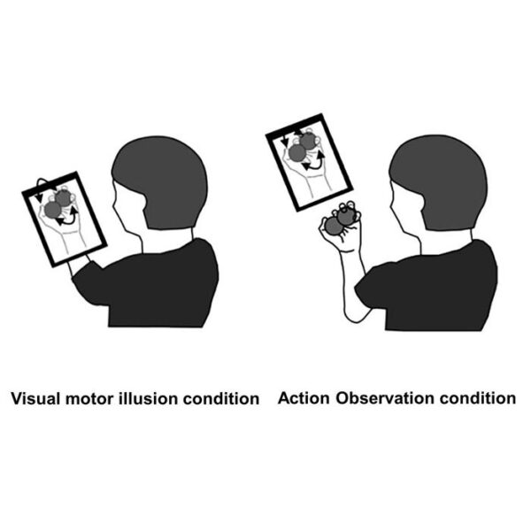
Visual-Motor Illusion (VMI) is an intriguing concept where you perceive your body moving despite being still. Imagine a tablet screen positioned in front of your motionless hand. The screen displays a video of your hand moving, creating the illusion that your hand is in motion, even though it’s not. This peculiar situation can be resolved by relocating the screen, making it just a regular video of hand movements to watch. Previous studies have shown differences in brain responses between VMI and action observation (AO), but the broader implications of VMI remained unclear.
Assistant Professor Katsuya Sakai and a team of scientists from Tokyo Metropolitan University conducted a study demonstrating that VMI can enhance both motor performance and the early stages of learning motor tasks. Volunteers were given a specific task involving rolling two metal balls around in one hand. Following initial tests, a visual aid displaying hands performing the exact action was introduced. One group had the aid positioned in front of their hand, inducing VMI, while the other group watched the video conventionally. Performance improvements were measured based on the number of successful ball rolls. While both groups showed enhancement, the VMI group exhibited greater improvement immediately after and one hour following the video presentation. This not only indicated performance enhancement but also highlighted improved early-stage learning that persisted over time.
To delve into the brain’s activity, the team employed functional near-infrared spectroscopy, a non-invasive method tracking brain activity using external probes. They observed notable differences between AO and VMI volunteers in brain regions linked to learning new movements. These changes persisted even an hour after the visual stimuli, aligning with the performance improvements observed in the task. This correlated with previous findings showing how VMI enhances connectivity in brain areas responsible for motor execution.
The researchers acknowledge the need for further exploration. While this study focused on healthy individuals, more investigation is necessary to evaluate mid to long-term motor performance effects. Nevertheless, these insights offer a unique approach to enhance motor skills and learning, potentially benefiting rehabilitation strategies for hemiplegic stroke patients and aiding the development of novel treatments.
This research received support from JSPS KAKENHI Grant Number 22K17569.


