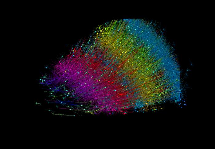
In a collaboration, researchers from Harvard University and Google have created the largest synaptic-resolution, 3D reconstruction of a piece of human brain to date. The cubic millimeter of brain tissue, which may seem small, contains an astounding 57,000 cells, 230 millimeters of blood vessels, and 150 million synapses, amounting to 1,400 terabytes of data.
The project, led by Jeff Lichtman, the Jeremy R. Knowles Professor of Molecular and Cellular Biology and newly appointed dean of science at Harvard, combines Lichtman’s electron microscopy imaging with Google’s AI algorithms to color-code and reconstruct the extremely complex wiring of mammal brains. The paper’s three co-first authors are former Harvard postdoctoral researcher Alexander Shapson-Coe, Michał Januszewski of Google Research, and Harvard postdoctoral researcher Daniel Berger.
Mapping the Brain: A Colossal Undertaking
The team’s ultimate goal, supported by the National Institutes of Health BRAIN Initiative, is to create a high-resolution map of a whole mouse brain’s neural wiring, which would entail about 1,000 times the amount of data they just produced from the human cortex fragment. “The word ‘fragment’ is ironic,” Lichtman said. “A terabyte is, for most people, gigantic, yet a fragment of a human brain – just a minuscule, teeny-weeny little bit of human brain – is still thousands of terabytes.”
Lichtman’s field, “connectomics,” seeks to create comprehensive catalogues of brain structure, down to individual cells and wiring. Such completed maps would light the way toward new insights into brain function and disease, about which scientists still know very little.
Discovering Rare Structures and Oddities
The latest map, published in Science, reveals never-before-seen details of brain structure, including a rare but powerful set of axons connected by up to 50 synapses. The team also noted oddities in the tissue, such as a small number of axons that formed extensive whorls. Since their sample was taken from a patient with epilepsy, they’re unsure if such unusual formations are pathological or simply rare.
Google’s state-of-the-art AI algorithms allow for reconstruction and mapping of brain tissue in three dimensions. The team has also developed a suite of publicly available tools researchers can use to examine and annotate the connectome. “Given the enormous investment put into this project, it was important to present the results in a way that anybody else can now go and benefit from them,” said Google Research collaborator Viren Jain.
Next, the team will tackle the mouse hippocampal formation, which is important to neuroscience for its role in memory and neurological disease. The analysis revealed that the under 4-minute milers lived nearly 5 years beyond their predicted life expectancy, on average, based on sex, age, year of birth, age at achievement, and nationality.
The researchers acknowledge that they didn’t have information on the lifelong exercise habits or other health behaviors of the 200 athletes included in the study, so they weren’t able to determine the precise relationship between lifelong exercise dose and longevity. Nevertheless, they say, “This finding challenges the upper ends of the U-shaped exercise hypothesis (as it relates to longevity) and, once again, reiterates the benefits of exercise on the lifespan, even at the levels of training required for elite performance.”
Using more than 1.4 petabytes of electron microscopy (EM) imaging data, the researchers have generated a nanoscale-resolution reconstruction of a millimeter-scale fragment of human cerebral cortex, providing an unprecedented view into the structural organization of brain tissue at the supracellular, cellular, and subcellular levels. The human brain is a vastly complex organ, and to date, little is known about its cellular microstructure, including the synaptic and neural circuits it supports. Disruption of these circuits is known to play a role in myriad brain disorders.
Shapson-Coe et al. generated a three-dimensional reconstruction of nearly every cell and process in the cubic millimeter sample and developed a freely available tool for visualizing and analyzing the vast dataset. Using the data, the authors discovered previously underappreciated aspects of the human temporal cortex, including the disproportionate number of glia over neurons and the existence of rare yet powerful axonal inputs that contain up to ~50 synapses. “Further studies using this resource may bring valuable insights into the mysteries of the human brain,” Shapson-Coe et al. write.
ScienceBlog.com has no paywalls, no sponsored content, and no agenda beyond getting the science right. Every story here is written to inform, not to impress an advertiser or push a point of view.
Good science journalism takes time — reading the papers, checking the claims, finding researchers who can put findings in context. We do that work because we think it matters.
If you find this site useful, consider supporting it with a donation. Even a few dollars a month helps keep the coverage independent and free for everyone.