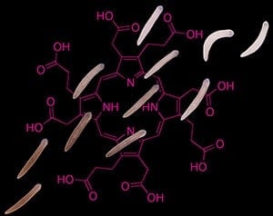
A type of flatworm could be a new weapon in the hunt for better ways to treat a group of diseases that can cause extreme sensitivity to light, facial hair growth, and hallucinations, according to a study published in the journal eLife.
Porphyrias are a group of rare metabolic disorders characterized by red and purple pigments accumulating in the body. With the accidental discovery that the skin color of the flatworm Schmidtea mediterranea (S. mediterranea) changes under prolonged exposure to sunlight, these animals could provide a new model for studying the diseases.
Faculty and student researchers from Keene State College, New Hampshire, show that the flatworm generates light-activated molecules called porphyrins in its skin pigment cells using the same biochemical pathway as that involved in human porphyrias. Their research developed from the unexpected observation by undergraduate students that the flatworms they were investigating changed color from brown to white when exposed to sunlight for several days. It could now lead to future drug development to meet patient demands for more effective treatments.
“Porphyrias are typically caused by inherited mutations that involve a buildup of porphyrins in various parts of the body. Although naturally created during the generation of heme, a substance required for oxygen transport in red blood cells, porphyrins can have toxic effects when their levels increase,” said Jason Pellettieri, corresponding author and Associate Professor of Biology at Keene State College. “For example, porphyrins deposited in the skin cause swelling, blistering, and lesions upon exposure to bright light. Neurological issues can also arise, ranging from anxiety and confusion to seizures or paralysis. These episodes, which can last for weeks, can be triggered by drugs, hormonal changes, and dieting or fasting.”
The porphyrins in S. mediterranea give rise to their normal skin pigmentation. When the animals are exposed to intense light for extended periods of time (a situation unlikely to occur in the wild), porphyrin production leads to pigment cell loss, changing the animals’ skin color to white. To investigate this photosensitivity, the researchers first tested infrared and ultraviolet lights on the flatworms, which had no effect on their skin. In contrast, intense visible light altered their coloring, with just over half of them developing one or more small tissue lesions.
“Our findings show that prolonged light exposure eliminates the flatworms’ pigment cells through a mechanism involving porphyrin-dependent photosensitization. The animals then repigment when they are no longer exposed to light,” explained lead author Bradford Stubenhaus.
During the course of the research, the team also noticed a positive relationship between how long the flatworms were fasted before light exposure and the extent of their photosensitivity.
To document this relationship, they starved the animals for one, seven, 14, or 30 days before exposing them to light. Depigmentation was strongly accelerated with starvation, and then reversed with a single feeding 24 hours before light exposure. These discoveries suggest that S. mediterranea could help identify new treatments for easing porphyrin-mediated photosensitivity or separating the relationship between dieting or fasting and the onset of disease symptoms.
“Although porphyrias are usually manageable diseases, reliance on the mainstay treatment, namely intravenous heme, or liver transplantation for more severe cases, can result in significant complications. There are also no approved preventative therapies for patients who suffer recurrent attacks,” says Pellettieri.
“Our studies show that flatworms such as S. mediterranea could potentially change this. They have recently emerged as a useful model for human disorders, including Usher syndrome – a genetic disorder that affects vision, hearing, and sometimes balance – and cystic kidney disease. We can now add porphyria to this growing list, with plans to use the animals to screen for novel therapies in the near future.”
Reference
The paper “Light-induced depigmentation in planarians models the pathophysiology of acute porphyrias” can be freely accessed online. Contents, including text, figures, and data, are free to reuse under a CC BY 4.0 license.


