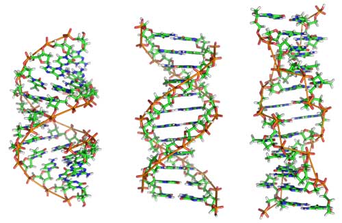
A new study could explain why DNA and not RNA, its older chemical cousin, is the main repository of genetic information. The DNA double helix is a more forgiving molecule that can contort itself into different shapes to absorb chemical damage to the basic building blocks — A, G, C and T — of genetic code. In contrast, when RNA is in the form of a double helix it is so rigid and unyielding that rather than accommodating damaged bases, it falls apart completely.
The research, published August 1, 2016 in the journal Nature Structural and Molecular Biology, underscores the dynamic nature of the DNA double helix, which is central to maintaining the stability of the genome and warding off ailments like cancer and aging. The finding will likely rewrite textbook coverage of the difference between the two purveyors of genetic information, DNA and RNA.
“There is an amazing complexity built into these simple beautiful structures, whole new layers or dimensions that we have been blinded to because we didn’t have the tools to see them, until now,” said Hashim M. Al-Hashimi, Ph.D., senior author of the study and professor of biochemistry at Duke University School of Medicine.
DNA’s famous double helix is often depicted as a spiral staircase, with two long strands twisted around each other and steps composed of four chemical building blocks called bases. Each of these bases contain rings of carbon, along with various configurations of nitrogen, oxygen, and hydrogen. The arrangement of these atoms allow G to pair with C and A to pair with T, like interlocking gears in an elegant machine.
When Watson and Crick published their model of the DNA double helix in 1953, they predicted exactly how these pairs would fit together. Yet other researchers struggled to provide evidence of these so-called Watson-Crick base pairs. Then in 1959, a biochemist named Karst Hoogsteen took a picture of an A-T base pair that had a slightly skewed geometry, with one base rotated 180 degrees relative to the other. Since then, both Watson-Crick and Hoogsteen base pairs have been observed in still images of DNA.
Five years ago, Al-Hashimi and his team showed that base pairs constantly morph back and forth between Watson-Crick and the Hoogsteen configurations in the DNA double helix. Al-Hashimi says that Hoogsteen base pairs typically show up when DNA is bound up by a protein or damaged by chemical insults. The DNA goes back to its more straightforward pairing when it is released from the protein or has repaired the damage to its bases.
“DNA seems to use these Hoogsteen base pairs to add another dimension to its structure, morphing into different shapes to achieve added functionality inside the cell,” said Al-Hashimi.
Al-Hashimi and his team wanted to know if the same phenomenon might also be occurring when RNA, the middleman between DNA and proteins, formed a double helix. Because these shifts in base pairing involve the movement of molecules at an atomic level, they are difficult to detect by conventional methods. Therefore, Al-Hashimi’s graduate student Huiqing Zhou used a sophisticated imaging technique known as NMR relaxation dispersion to visualize these tiny changes. First, she designed two model double helices — one made of DNA and one made of RNA. Then, she used the NMR technique to track the flipping of individual G and A bases that make up the spiraling steps, pairing up according to Watson-Crick or Hoogsteen rules.
Prior studies indicated that at any given time, one percent of the bases in the DNA double helix were morphing into Hoogsteen base pairs. But when Zhou looked at the corresponding RNA double helix, she found absolutely no detectable movement; the base pairs were all frozen in place, stuck in the Watson-Crick configuration.
The researchers wondered if their model of RNA was an unusual exception or anomaly, so they designed a wide range of RNA molecules and tested them under a wide variety of conditions, but still none appeared to contort into the Hoogsteen configuration. They were concerned that the RNA might actually be forming Hoogsteen base pairs, but that they were happening so quickly that they weren’t able to catch them in the act. Zhou added a chemical known as a methyl group to a specific spot on the bases to block Watson-Crick base pairing, so the RNA would be trapped in the Hoogsteen configuration. She was surprised to find that rather than connecting through Hoogsteen base pairs, the two strands of RNA came apart near the damage site.
“In DNA this modification is a form of damage, and it can readily be absorbed by flipping the base and forming a Hoogsteen base pair. In contrast, the same modification severely disrupts the double helical structure of RNA,” said Zhou, who is lead author of the study.
The team believes that RNA doesn’t form Hoogsteen base pairs because its double helical structure (known as A-form) is more compressed than DNA’s (B-form) structure. As a result, RNA can’t flip one base without hitting another, or without moving around atoms, which would tear apart the helix.
“For something as fundamental as the double helix, it is amazing that we are discovering these basic properties so late in the game,” said Al-Hashimi. “We need to continue to zoom in to obtain a deeper understanding regarding these basic molecules of life.”
The research was supported by grants from the National Institutes of Health (R01GM089846 and 5P50GM103297) and Austrian Science Fund (P26550 and P28725).
CITATION: “m1A and m1G Potently Disrupt A-RNA Structure Due to the Intrinsic Instability of Hoogsteen Base Pairs,”Huiqing Zhou, Isaac J. Kimsey, Evgenia N. Niklova, Bharathwaj Sathyamoorthy, Gianmarc Grazioli, James McSally, Tianyu Bai, Christoph H. Wunderlich, Christoph Kreutz, Ioan Andricioaei, and Hashim M. Al-Hashimi. Nature Structural and Molecular Biology, Aug. 1, 2016. https://dx.doi.org/10.1038/nsmb.3270.


