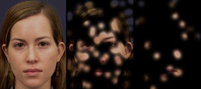
Responding to faces is a critical tool for social interactions between humans. Without the ability to read faces and their expressions, it would be hard to tell friends from strangers upon first glance, let alone a sad person from a happy one. Now, neuroscientists from the California Institute of Technology (Caltech), with the help of collaborators at Huntington Memorial Hospital in Pasadena and Cedars-Sinai Medical Center in Los Angeles, have discovered a novel response to human faces by looking at recordings from brain cells in neurosurgical patients.
The finding, described in the journal Current Biology, provides the first description of neurons that respond strongly when the patient sees an entire face, but respond much less to a face in which only a very small region has been erased.
“The finding really surprised us,” says Ueli Rutishauser, first author on the paper, a former postdoctoral fellow at Caltech, and now a visitor in the Division of Biology. “Here you have neurons that respond well to seeing pictures of whole faces, but when you show them only parts of faces, they actually respond less and less the more of the face you show. That just seems counterintuitive.”
The neurons are located in a brain region called the amygdala, which has long been known to be important for the processing of emotions. However, the study results strengthen a growing belief among researchers that the amygdala has also a more general role in the processing of, and learning about, social stimuli such as faces.
Other researchers have described the amygdala’s neuronal response to faces before, but this dramatic selectivity—which requires the face to be whole in order to elicit a response—is a new insight.
“Our interpretation of this initially puzzling effect is that the brain cares about representing the entire face, and needs to be highly sensitive to anything wrong with the face, like a part missing,” explains Ralph Adolphs, senior author on the study and Bren Professor of Psychology and Neuroscience and professor of biology at Caltech. “This is probably an important mechanism to ensure that we do not mistake one person for another and to help us keep track of many individuals.”
The team recorded brain-cell responses in human participants who were awaiting surgery for drug-resistant epileptic seizures. As part of the preparation for surgery, the patients had electrodes implanted in their medial temporal lobes, the area of the brain where the amygdala is located. By using special clinical electrodes that have very small wires inserted, the researchers were able to observe the firings of individual neurons as participants looked at images of whole faces and partially revealed faces. The voluntary participants provided the researchers with a unique and very rare opportunity to measure responses from single neurons through the implanted depth electrodes, says Rutishauser.
“This is really a dream collaboration for basic research scientists,” he says. “At Caltech, we are very fortunate to have several nearby hospitals at which the neurosurgeons are interested in such collaborative medical research.”
The team plans to continue their studies by looking at how the same neurons respond to emotional stimuli. This future work, combined with the present study results, could be highly valuable for understanding a variety of psychiatric diseases in which this region of the brain is thought to function abnormally, such as mood disorders and autism.
Other Caltech authors on the paper are Oana Tudusciuc, a postdoctoral scholar in neuroscience and psychology, and Dirk Neumann, visiting associate in biology. Medical collaborators on the study include Adam Mamelak, a neurosurgeon at Cedars-Sinai; and Huntington Memorial Hospital’s Christopher Heller, neurosurgeon; Ian Ross, neurosurgeon; Linda Philpott, neuropsychologist; and William Sutherling, medical director of the Epilepsy and Brain Mapping Program. The work in the paper “Neuronal responses selective for whole faces in the human amygdala,” was supported by the National Science Foundation, the Pfeiffer Family Foundation, and the Gordon and Betty Moore Foundation.


