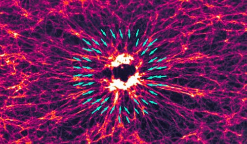
Mechanical forces are a critical part of essential cellular behaviors, from muscle contraction to cell division. Exactly how they generate those forces, however, isn’t entirely understood. An artificial cellular environment and a photosensitive drug have now given researchers a clearer picture of the process and potentially clues to how cancer cells behave.
The results of that study, led by Michael Murrell, a Yale assistant professor of biomedical engineering, and his collaborators at the University College of London and Purdue University, are published today in Nature Communications.
How the organization of cells’ internal machine — the cytoskeleton — affects the forces they generate is a longstanding question, particularly regarding the differences between the cytoskeleton architectures in muscle cells and non-muscle cells.
In muscle cells, the cytoskeleton is highly organized; filaments of actin and myosin — two of the primary proteins that generate the mechanical forces in animal cells — are neatly aligned. The myosin acts as a motor, binding to the actin and then pulling it and releasing it. The actin filaments slide past the myosin, which repeats the process. This produces tensile forces, called “contracts,” that shorten the cytoskeleton.
These tensile forces are used for “pulling” on the extracellular matrix, which promotes cell migration. The contractions are described as “telescopic” — that is, the shortening increases in speed with the size (length) of the cytoskeletal machinery. Researchers have long assumed this to be the direct result of the organized structure of the cells’ architectures.
But the cytoskeleton of non-muscle cells appears to be very disordered. Actin filaments, known as F-actin, have varying lengths and no net orientation or polarity. And yet, contractility still happens.
Understanding these forces could help explain the behavior of cancer cells. “Metastasis is a very mechanical process,” said Murrell, senior author of the study and a member of the Yale Systems Biology Institute at Yale’s West Campus. “A tumor cell will break free from its local environment and move into the extracellular space. During this process, there are dramatic changes in the organization of F-actin and the cell’s ability to generate and transmit force. Understanding how organization and architecture relate to contractility would be very valuable.”
To better study the behavior of actin and myosin, Murrell’s research group purified these proteins from non-muscle cells and reconstructed them into a biomimetic model of the cytoskeleton. To control the activity, they used blebbistatin, a drug that inhibits myosin. For many researchers, the drug is frustrating because it is photosensitive and becomes inactive when blue light is used for imaging. For Murrell’s purposes, though, this “flaw” was perfect. By precisely directing the light, researchers could activate the myosin when and where they wanted, while keeping it stabilized (or inactive) in other parts of the cytoskeleton.
“It allows us to control contractility in space and time,” Murrell said. “We activated these regions of different sizes and studied the velocity of the contractions.”
The researchers found that the larger the area in which the cytoskeleton was activated, the higher the velocity of contractions generated by the actomyosin. This indicates that it’s the active and viscoelastic nature of the material — not the order seen in muscle cells — causing the telescopic contractility, they said.


