In our increasingly health-conscious society, a new fad diet seems to pop up every few years. Atkins, Zone, Ketogenic, Vegetarian, Vegan, South Beach, Raw – with so many choices and scientific evidence to back each, it’s hard to know what’s healthy and what’s not. One message, however, has remained throughout: saturated fats are bad.
A new Columbia University study reveals why.
While doctors, nutritionists and researchers have known for a long time that saturated fats contribute to some of the leading causes of death in the United States, they haven’t been able to determine how or why excess saturated fats, such as those released from lard, are toxic to cells and cause a wide variety of lipid-related diseases, while unsaturated fats, such as those from fish and olive oil, can be protective.
To find answers, Columbia researchers developed a new microscopy technique that allows for the direct tracking of fatty acids after they’ve been absorbed into living cells. The technique involves replacing hydrogen atoms on fatty acids with their isotope, deuterium, without changing their physicochemical properties and behavior like traditional strategies do. By making the switch, all molecules made from fatty acids can be observed inside living cells by an advanced imaging technique called stimulated Raman scattering (SRS) microscopy.
What the researchers found using this technique could have significant impact on both the understanding and treatment of obesity, diabetes and cardiovascular disease.
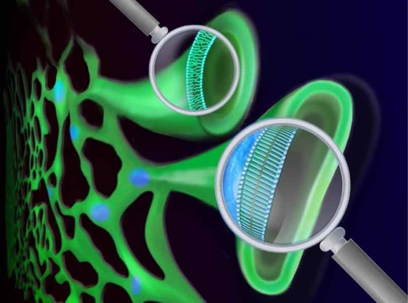
Published online December 1st in Proceedings of the National Academy of Sciences (PNAS), the team reports that the cellular process of building the cell membrane from saturated fatty acids results in patches of hardened membrane in which molecules are “frozen.” Under healthy conditions, this membrane should be flexible and the molecules fluidic.
The researchers explained that the stiff, straight, long chains of saturated fatty acids rigidify the lipid molecules and cause them to separate from the rest of the cell’s membrane. Under their microscope, the team observed that those lipid molecules then accumulate in tightly-packed “islands,” or clusters, that don’t move much – a state they call “solid-like.” As more saturated fatty acids enter the cell, those islands grow in size, creating increasing inelasticity of the membrane and gradually damaging the entire cell.
“For a long time, we believed that all cell membrane is liquid-like, allowing embedded proteins to change their shape and perform reactions,” said Principal Investigator Wei Min, a professor of chemistry. “Solid-like membrane was hardly observed in living mammalian cells before. What we saw was quite different and surprising.”
Lipid molecules made from unsaturated fatty acids on the other hand bear a kink in their chains, Min said, which makes it impossible for these lipid molecules to align closely with each other as saturated ones do. They continue to move around freely rather than forming stationary clusters. In their movement, these molecules can jostle and slide in between the tightly-packed saturated fatty acid chains.
“We found that adding unsaturated fatty acids could ‘melt’ the membrane islands frozen by saturated fatty acids,” said First Author Yihui Shen, a graduate student in Min’s lab. This new mechanism, she said, can partly explain the beneficial effect of unsaturated fatty acids and how unsaturated fats like those from fish oil can be protective in some lipid disorders.
The study represents the first time researchers were able to visualize the distribution and dynamics of fatty acids in such detail inside living cells, Shen added, and it revealed a previously unknown toxic physical state of the saturated lipid accumulation inside cellular membranes.
“The behavior of saturated fatty acids once they’ve entered cells contributes to major and often deadly diseases,” Min said. “Visualizing how fatty acids are contributing to lipid metabolic disease gives us the direct physical information we need to begin looking for effective ways to treat them. Perhaps, for example, we can find a way to block the toxic lipid accumulation. We’re excited. This finding has the potential to really impact public health, especially for lipid related diseases.”


The study doesn’t mention which saturated fats were tested, and implies that shorter and longer chain fatty acids all produce similar results. It would be interesting to compare the effect on cell membranes between stearic acid (C18) and caprylic acid (C8).
Now explain why the brain is almost 50% saturated fat… This shows how much science does not know..