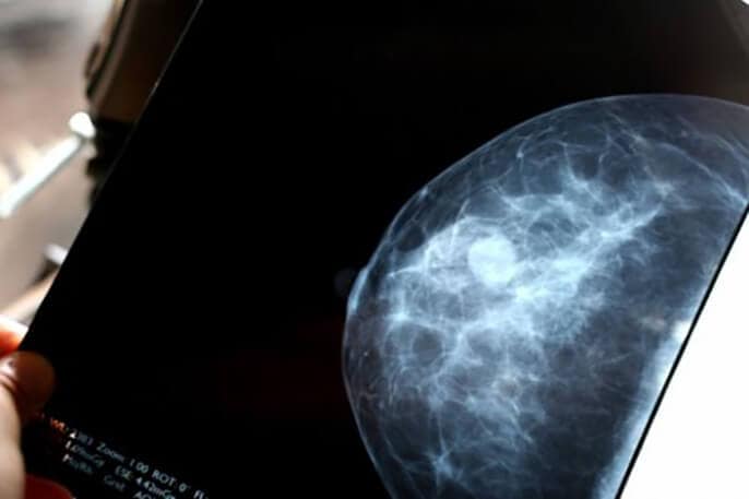
Automated breast-density evaluation was just as accurate in predicting women’s risk of breast cancer, found and not found by mammography, as subjective evaluation done by radiologists, in a study led by researchers at UC San Francisco and Mayo Clinic.
Both assessment methods were equally accurate in predicting both the risk of cancer detected through mammography screening and the risk of interval invasive cancer – that is, cancer diagnosed within a year of a negative mammography result. Both methods predicted interval cancer more strongly than screen-detected cancer. The study was published May 1, 2018, in the Annals of Internal Medicine.
Breast density can increase tumor aggressiveness, as well as mask the presence of tumors in mammograms, explained UCSF Professor of Medicine Karla Kerlikowske, MD, who led the study with Mayo Clinic Professor of Epidemiology Celine Vachon, PhD. “This means that women with dense breasts are more likely to be diagnosed with advanced-stage breast cancers, especially those that are interval cancers, because their cancers are more likely to remain undetected for longer.”
“These findings demonstrate that breast-density evaluation can be done with equal accuracy by either a radiologist or an automated system,” said Kerlikowske. “They also show the potential value of a reproducible automated evaluation in helping identify women with dense breasts who are at higher risk of aggressive tumors, and thus more likely to be candidates for supplemental screening.”
Another important result of the study was that both automated and clinical breast density assessment methods were equally accurate at predicting both screen-detected and interval cancers when breast density was assessed within 5 years of cancer diagnosis.
“These findings have implications for breast cancer risk models, in particular those that predict risk of the more aggressive interval cancer,” said Vachon, the paper’s senior author. “Breast density measures can be used to inform a woman’s risk of screen and/or interval detected cancers up to five years after assessment.”
Thirty states have laws requiring that women receive notification of their breast density, which is graded on the standard four-category Breast Imaging Reporting and Data System (BI-RADS) scale: a. almost entirely fatty; b. scattered fibroglandular densities; c. heterogeneously dense; and d. extremely dense. “Women who are accurately identified to have dense breasts and be at high risk of an interval cancer are more likely to have appropriate discussions of whether supplemental imaging is right for them,” said Kerlikowske.
The study, the largest of its kind to date, included 1,609 women with cancer detected within a year of a positive mammography result, 351 women with interval invasive cancer, and 4,409 control participants matched by age, race, state of residence, screening date and mammography machine. The participants came from screening practices in San Francisco and Rochester, Minn.
The researchers found that women with an automated BI-RADS assessment of extremely dense breasts had a 5.65 times higher risk of interval cancer and a 1.43 times higher risk of screen-detected cancer than women with scattered fibroglandular densities, the most common density category in average-risk women.
“There have been concerns raised about the reliability of BI-RADS breast density measures, since an assessment might vary for an individual woman depending on the radiologist and the mammogram,” observed Kerlikowske, who was first author on the paper. “Automated assessments, which are done by computer algorithm, are more reproducible and less subjective. Therefore, they could reduce variation and alleviate the sense of subjectivity and inconsistency.”


