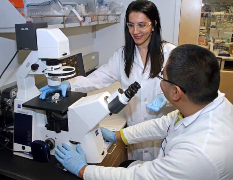
Origami – the Japanese art of folding paper into shapes and figures – dates back to the sixth century. At UMass Lowell, it is inspiring researchers as they develop a 21st century solution to the shortage of tissue and organ donors.
Gulden Camci-Unal, an assistant professor of chemical engineering, and her team of student researchers are designing new biomaterials that could someday be used to repair, replace or regenerate skin, bone, cartilage, heart valves, heart muscle and blood vessels, and in other applications.
Using origami as inspiration, Camci-Unal and her team are using plain paper to create centimeter-scale scaffoldings where the cells can grow and then applying microfabrication techniques to generate new biomaterials known as tissue mimetics.
“Paper is a low-cost, widely available and extremely flexible material that can be easily fabricated into three-dimensional structures of various shapes, sizes and configurations,” said Camci-Unal.
The team uses origami-folded paper to grow bone cells, called osteoblasts, which produce the matrix that gets deposited with minerals to form bone. The paper can then be implanted to treat patients, including those suffering from damage caused by disease, degeneration or trauma, or bone defects such as irregular sizes and shapes.
So far, the team’s research indicates the implants are biocompatible – that is, they are not expected to be rejected by the body’s immune system, according to Camci-Unal.
The team is also using their paper-based research to learn about the behavior of lung cancer cells.
“Tumor biopsies from patients can be grown in our system and then these cells can be exposed to different chemotherapy drugs or radiation doses to find out which specific treatment would work best for the patient,” said Camci-Unal, who joined UMass Lowell’s faculty in 2016.
The Camci-Unal research group is also developing low-cost, paper-based platforms for point-of-care disease diagnostics. Their biosensors can be used to determine if a patient has a specific disease.
In addition to paper, Camci-Unal and her team are using hydrogels – flexible, squishy materials that resemble Jell-O and are made mostly from water – in tissue-related research for a range for a variety of uses, including wound care.
“Their physical, chemical and biological properties can be tailored to fit in various tissue engineering applications,” she said. “However, hydrogels have relatively weak mechanical properties, so they tend to be not as easy to handle and manipulate when they are in large, very thin sheets.”
By combining hydrogels laden with cells with sheets of paper, Camci-Unal is able to create sufficiently strong support structures that can be used for tissue engineering. Other researchers have developed 3-D structures from synthetic materials like polymers, ceramics and metals to culture cells, but most of those traditional materials do not closely resemble the surroundings in native tissues, she explained.
“Our team recently started to get involved in wound-healing research, too. Our ultimate goal is to improve human health and the quality of life,” Camci-Unal said.
Camci-Unal and three of her students – biology major Kierra Walsh of Billerica and Xinchen Wu and Sanika Suvarnapathaki, both of Lowell and Ph.D. students in biomedical engineering and biotechnology – discussed the use of paper-based, 3-D platforms for cell cultures and other biomedical applications in a Jan. 24 article in MRS Communications, a peer-reviewed academic journal used by researchers worldwide for the rapid dissemination of breakthroughs in materials science.


