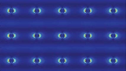
Researchers have developed a tiny nanolaser that can function inside living tissues without harming them. Just 50 to 150 nanometers thick, the laser is about 1/1,000th the thickness of a single human hair. At this size, the laser can fit and function inside living tissues, with the potential to sense disease biomarkers or perhaps treat deep-brain neurological disorders, such as epilepsy.
Developed by NSF-funded researchers at Northwestern and Columbia Universities, the nanolaser shows promise for imaging in living tissues. Not only is the laser made mostly of glass, which is intrinsically biocompatible, but it can also be excited with longer wavelengths of light and emit at shorter wavelengths.
“Longer wavelengths of light are needed for bioimaging because they can penetrate farther into tissues than visible wavelength photons,” said Northwestern’s Teri Odom, who co-led the research. “But shorter wavelengths of light are often desirable at those same deep areas. We have designed a system that can deliver visible laser light at penetration depths accessible to longer wavelengths.”
Odom co-led the work with P. James Schuck at Columbia University’s School of Engineering.
The nanolaser also can operate in extremely confined spaces, including quantum circuits and microprocessors for ultra-fast and low-power electronics.
The paper was published in the journal Nature Materials.
While many applications require increasingly small lasers, researchers continually run into the same roadblock: Nanolasers tend to be much less efficient than their macroscopic counterparts. And these lasers typically need shorter wavelengths, such as ultraviolet light, to power them.
Odom, Schuck and their teams were able to achieve a nanolaser platform that solves these issues by using photon upconversion. In upconversion, low-energy photons are absorbed and converted into one photon with higher energy.
The research was funded by NSF’s Division of Materials Research.


