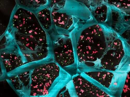
To many of us, cells are the building blocks of life, akin to bricks or Legos. But to biologist Regan Moore, a former Ph.D. student in Dan Kiehart’s lab at Duke, cells are so much more: they’re busy construction sites, machinery and materials moving about to build and shape the body. And now, new live imaging techniques make it possible to watch some of the nano-scale construction in action.
In time-lapse videos published this month, Moore, Kiehart and colleagues were able to peer inside cells as they filled a hole in the back of a developing fruit fly, a crucial step in the fly’s development into a larva. The process is coordinated with help from a thin mesh of protein fibers just under the cell surface, each one 10,000 times finer than a human hair. The fibers help the cells hold their shape, “kind of like rebar in concrete,” Moore said.

But unlike rebar, she added, “they’re constantly moving and changing.” Normally, features like these are too small and quick to see with conventional microscopes, which can only take a few images a second or are too out-of-focus. So Kiehart’s team used a technique called super-resolution fluorescence microscopy to track individual fibers with nanoscale resolution.
By watching the “rebar of the cell” at work during this hole-closing process in fruit flies, the researchers hope to better understand wound healing in humans, and what goes awry for children with birth defects such as cleft lip and spina bifida.
LEARN MORE: “Super-resolution microscopy reveals actomyosin dynamics in medioapical arrays,” Regan P. Moore, Stephanie M. Fogerson, U. Serdar Tulu, Jason W. Yu, Amanda H. Cox, Melissa A. Sican, Dong Li, Wesley R. Legant, Aubrey V. Weigel, Janice M. Crawford, Eric Betzig, and Daniel P. Kiehart. Molecular Biology of the Cell, July 15, 2022. DOI: 10.1091/mbc.E21-11-0537


