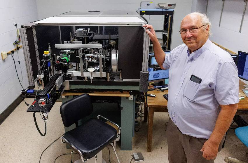
Indiana University scientists are developing advanced ophthalmoscopes that could detect diabetes, heart disease, and Alzheimer’s through simple eye scans. This research, part of the NIH’s new “oculomics” initiative, aims to revolutionize early disease detection.
Summary: Indiana University researchers receive $4.8 million NIH grant to develop next-generation eye scanning technology capable of detecting early signs of major diseases. The project combines advanced optics with artificial intelligence to create a powerful, non-invasive diagnostic tool.
Estimated reading time: 5 minutes
The human eye, long known as a window to the soul, may soon become a window to our overall health. Researchers at Indiana University are pioneering the development of advanced eye-scanning technology that could detect early signs of major diseases like diabetes, heart disease, and Alzheimer’s through a simple, non-invasive exam.
Stephen A. Burns, a professor at the IU School of Optometry, leads this groundbreaking project as part of the National Institutes of Health’s new Venture Program Oculomics Initiative. The three-year, $4.8 million award supports the emerging field of “oculomics,” which uses the eye as a lens to observe diseases affecting the entire body.
The Eye as a Health Monitor
“This research is about using the eye as a window on health,” Burns said, noting that the retina is the only directly observable part of the central nervous system. “We want to give health care providers the clearest view they can hope to get into the body, non-invasively.”
The project builds on Burns’ previous work in applying adaptive optics to eye observation. This technology, originally developed by astronomers to eliminate atmospheric distortions in telescopes, allows for incredibly detailed views of the human eye.
The ophthalmoscope in Burns’ lab can observe the back of the human eye at a resolution of two microns – small enough to show the real-time movement of individual red blood cells inside the eye’s blood vessels. This level of detail has already allowed researchers to identify biomarkers for diabetes and hypertension in the walls of these vessels.
Integrating Technologies for Broader Detection
The NIH-funded project will combine the work of several research teams to create a single, powerful diagnostic tool. Researchers from Northwestern University and Mount Sinai have used similar technology to observe cells both inside and outside blood vessels, including the distinctive crescent-shaped cells found in sickle cell anemia. Stanford University researchers have applied adaptive optics to improve observation of the eye’s photoreceptors.
By integrating these technologies and applying state-of-the-art machine learning and AI, the team hopes to expand the device’s capabilities to detect early signs of heart disease and Alzheimer’s disease.
“There’s growing evidence of a strong retinal vascular component to Alzheimer’s disease,” Burns explained. “You can currently see the signs with PET scans, which require large, multimillion-dollar instruments. If we can see the same signs with an eye scan, it’s a lot less invasive and a lot less costly.”
The Role of Artificial Intelligence
Eleftherios Garyfallidis, an associate professor of intelligent systems engineering at IU’s Luddy School of Informatics, Computing and Engineering, will lead the development of machine learning and AI methods for interpreting the devices’ results. This could dramatically reduce diagnosis time from days to minutes by eliminating the need for human analysis of the imagery.
The project will unfold in three phases over the next three years. First, the research teams will align their instruments to the same level of sensitivity. Next, they will validate the data to confirm that the new instruments’ readings align with earlier versions of the technology and that the AI system’s interpretations match those of human analysts. Finally, the device will be tested on clinical volunteers, with much of IU’s data coming from individuals recruited through the Atwater Eye Care Center.
Potential Impact and Challenges
The potential impact of this technology is significant. “Up to 80 percent of the population over the age of 60 has at least one health issue that may be detectable in the eye with our technology,” Burns noted.
However, challenges remain. “Our challenge now is selectivity and specificity,” Burns said. “We need to show that we can detect the differences between conditions, to quickly and accurately interpret the signs of the various diseases we’re focusing on.”
The ultimate goal is to advance the technology until it is ready for widespread use in routine eye exams. If successful, this research could transform early disease detection, making it possible to spot the warning signs of major health issues with a simple trip to the optometrist.
Quiz: Test Your Knowledge
- What is the resolution at which the ophthalmoscope in Burns’ lab can observe the back of the human eye?
- Which disease’s distinctive cells have researchers observed using this technology?
- What is the ultimate goal for the application of this technology?
Answers:
- Two microns
- Sickle cell anemia
- To be used in routine eye exams for widespread disease detection
Glossary of Terms
- Oculomics: An emerging field that uses the eye as a lens to observe diseases affecting the entire body.
- Ophthalmoscope: An instrument used to observe the interior of the eye.
- Adaptive Optics: A technology originally developed for astronomy, now applied to improve observations of the human eye.
- Retina: The light-sensitive layer at the back of the eye, and the only directly observable part of the central nervous system.
- Biomarkers: Measurable indicators of the presence or severity of a disease.
- Photoreceptors: Specialized neurons in the retina that respond to light.
Enjoy this story? Get our newsletter! https://scienceblog.substack.com/


