Summary: Harvard researchers have developed a groundbreaking technique to detect early signs of Parkinson’s disease through blood analysis. Their innovative method examines tiny cellular packages called extracellular vesicles, potentially enabling diagnosis years before symptoms appear. This advancement could revolutionize how we diagnose and treat brain disorders.
Journal: Proceedings of the National Academy of Sciences, October 30, 2024, DOI: 10.1073/pnas.2408949121
Reading time: 5 minutes
The Race for Earlier Detection
Brain disorders like Parkinson’s disease begin their silent progression long before the first tremor or other visible symptoms appear. Currently, definitive diagnosis requires examining brain tissue – something that can only be done after death. But that might be about to change.
Researchers at Harvard’s Wyss Institute have developed a new technique that could revolutionize how we detect brain diseases through a simple blood test. This approach focuses on analyzing tiny cellular packages called extracellular vesicles (EVs) that float in our blood.
A Technical Breakthrough
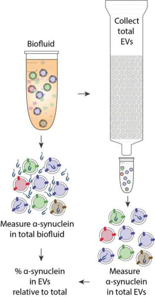
The research team, led by David Walt, Ph.D., has solved a crucial problem that has long puzzled scientists. They developed a method to determine exactly which disease markers are carried inside these cellular packages, rather than just stuck to their surface.
As Walt explains: “Research on EVs in our and other groups over the last few decades has steadily advanced our understanding of their complex biology and molecular composition. Yet, the isolation of pure tissue-specific EVs from body fluids like blood… and validating and quantifying their true contents with precise measurements still present formidable technical challenges.”
Promising Results for Patients
The team tested their method on blood samples from patients with Parkinson’s disease and a related condition called Lewy Body Dementia. They found that certain forms of the disease-related protein α-synuclein were two to three times more concentrated inside the vesicles compared to outside them.
Donald Ingber, Wyss Founding Director, highlights the significance: “The work by David Walt’s team presents a technological tour-de-force that brings us closer and closer to a next-generation diagnostic platform with extraordinary potential. At this point, we are not far from using these extremely rich and telling cell-derived vesicles as a window to peak into the brains of patients without requiring surgery.”
Technical Terms Glossary
Extracellular Vesicles (EVs): Tiny membrane-bound sacs released by cells into body fluids, containing various molecules from their origin cells.
α-synuclein: A protein that becomes modified during Parkinson’s disease progression and serves as a key biomarker.
Liquid Biopsy: A medical test done on blood or other body fluids to look for signs of disease, offering a less invasive alternative to tissue sampling.
Biomarker: A measurable substance in the body that indicates the presence of a disease or condition.
Test Your Knowledge
1. Why is current Parkinson’s disease diagnosis limited?
Answer: Brain tissue can only be examined posthumously for definitive diagnosis.
2. What are extracellular vesicles?
Answer: Tiny membrane-bound sacs released by cells into body fluids.
3. How much more concentrated was the modified α-synuclein inside vesicles compared to outside?
Answer: Two to three times more concentrated.
4. What is the potential impact of this research?
Answer: It could enable early diagnosis of brain disorders through simple blood tests.
Enjoy this story? Get our newsletter! https://scienceblog.substack.com/


