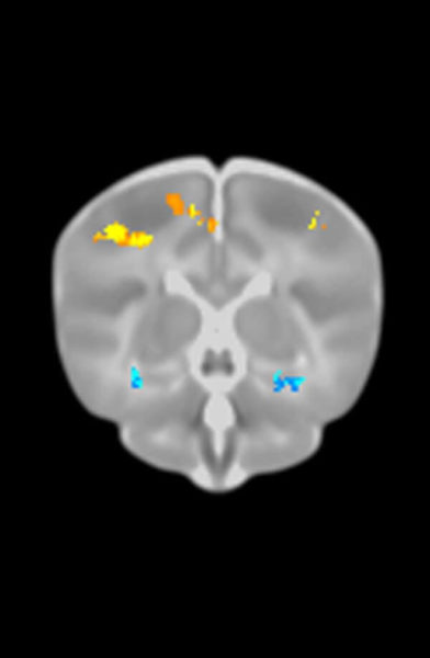
The rates of regional brain loss and cognitive decline caused by aging and the early stages of Alzheimer’s disease (AD) are higher for women and for people with a key genetic risk factor for AD, say researchers at the University of California, San Diego School of Medicine in a study published online July 4 in the American Journal of Neuroradiology.
The linkage between APOE ε4 – which codes for a protein involved in binding lipids or fats in the lymphatic and circulatory systems – was already documented as the strongest known genetic risk factor for sporadic AD, the most common form of the disease. But the connection between the sex of a person and AD has been less-well recognized, according to the UC San Diego scientists.
“APOE ε4 has been known to lower the age of onset and increase the risk of getting the disease,” said the study’s first author Dominic Holland, PhD, a researcher in the Department of Neurosciences at UC San Diego School of Medicine. “Previously we showed that the lower the age, the higher the rates of decline in AD. So it was important to examine the differential effects of age and APOE ε4 on rates of decline, and to do this across the diagnostic spectrum for multiple clinical measures and brain regions, which had not been done before.”
The scientists evaluated 688 men and women over the age of 65 participating in the Alzheimer’s Disease Neuroimaging Initiative, a longitudinal, multi-institution study to track the progression of AD and its effects upon the structures and functions of the brain. They found that women with mild cognitive impairment (a condition precursory to AD diagnosis) experienced higher rates of cognitive decline than men; and that all women, regardless of whether or not they showed signs of dementia, experienced greater regional brain loss over time than did men. The magnitude of the sex effect was as large as that of the APOE ε4 allele.
“Assuming larger population-based samples reflect the higher rates of decline for women than men, the question becomes what is so different about women,” said Holland. Hormonal differences or change seems an obvious place to start, but Holland said this is largely unknown territory – at least regarding AD.
“Another important finding of this study is that men and women did not differ in the level of biomarkers of Alzheimer’s disease pathology,” said co-author Linda McEvoy, PhD, an associate professor in the UCSD Department of Radiology. “This suggests that brain volume loss in women may also be caused by factors other than Alzheimer’s disease, or that in women, these pathologies are more toxic. We clearly need more research on how an individual’s sex affects AD pathogenesis.”
Holland acknowledged that the paper likely raises more questions than it answers. “There are many factors that may affect the sex differences we observed, such as whether the women in this study may have had higher rates of diabetes or insulin resistance than the men. We also do not know how the use of hormone replacement therapy, reproductive history or years since menopause may have affected these differences. All these issues need to be examined. There is no prevailing theory.”
But he said that just as APOE ε4 status identifies individuals at greater risk of AD, the sex of a person might prove an important determinant in future treatment as well. Currently, there is no cure for AD or any existing therapies that slow or stop disease progression.
“The biggest impact might be down the road when disease-modifying therapies become available,” said Holland. “What works best for men might not work best for women. The same may be true for ε4 carriers versus non-carriers.”
He added that results also feed back into clinical trial design. The sex-makeup of the sample will affect the rates of decline for both natural progression (the placebo component) and, likely, the degree of disease modification in participants receiving therapy. So a sex-based sub-analysis might be appropriate.
“Additionally, in clinical practice it may be important to expect higher rates of decline for women patients, to help anticipate when stages of decline that significantly alter quality of life would be reached.”


