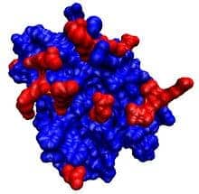
Researchers at UC San Francisco have discovered that endostatin, a protein that once aroused intense interest as a possible cancer treatment, plays a key role in the stable functioning of the nervous system.
A substance that occurs naturally in the body, endostatin potently blocks the formation of new blood vessels. In studies in mice in the late 1990s, endostatin treatment virtually eliminated cancer by shutting down the blood supply to tumors, but subsequent human clinical trials proved disappointing.
“It was a very big surprise” to find that endostatin, through some other mechanism, helps to maintain the proper workings of synapses, the sites where communication between nerve cells takes place, said Graeme W. Davis, PhD, Hertzstein Distinguished Professor of Medicine in the Department of Biochemistry and Biophysics at UCSF and senior author of the new study. “Endostatin was not on our radar.”
The findings were reported online July 24 in the journal Neuron.
Synapses are continually shaped and reshaped by experience, a phenomenon known as plasticity. But for those changes to be meaningful, said Davis, they must take place against a stable background, which paradoxically requires another form of change that he and colleagues call “homeostatic plasticity.” Just as we change our pace, slowing down or speeding up, to keep abreast of a running partner, neurons adjust aspects of their function at synapses to compensate for changes in their synaptic partners brought on by aging, illness, or other factors.
In an example of homeostatic plasticity, in the neuromuscular disease myasthenia gravis, as muscle cells become less responsive to the neurotransmitter acetylcholine, nerve cells ramp up their secretion of the neurotransmitter to keep the system in balance for as long as possible. Some researchers believe that in other disorders, including autism and schizophrenia, a failure in such homeostatic mechanisms keeps synapses from functioning properly.
In previous research Davis noticed that applying a toxin to a muscle cell in the fruit fly Drosophila melanogaster triggers homeostatic plasticity in the neuron that forms a synapse on that muscle cell: the neuron–which is called presynaptic, because it is “before” the synapse with the muscle cell–reliably releases more neurotransmitter, just as happens when muscle cells begin to malfunction in myasthenia gravis.
Davis has since built on this model of homeostatic plasticity by painstakingly knocking out Drosophila genes one by one and recording from presynaptic neurons to see which genes are necessary for the homeostatic response, because it is these genes that may be compromised in diseases affecting the process.
“So far we’ve tested about 1,000 genes this way, which has entailed close to 10,000 recordings,” Davis said.
Using this technique Davis and colleagues observed at one point that knocking out a gene called multiplexin significantly hampered homeostatic plasticity in presynaptic neurons. But because that gene helps to form a structural protein known as collagen—which in humans is a component of ligaments, tendons, and cartilage—the finding wasn’t immediately considered relevant to synaptic function.
The team learned that the multiplexin protein can be snipped by an enzyme to produce endostatin, so in experiments led by postdoctoral fellow Tingting Wang, PhD, they tested whether endostatin might play a role in homeostatic plasticity.
“Nobody picked up multiplexin to work on for a couple of years, because we didn’t think a collagen could be that interesting,” Davis said. “Then, when a new postdoc, Tingting Wang, came to the lab, we started thinking about it harder.”
When the group genetically deleted the portion of Drosophila multiplexin that forms endostatin, presynaptic neurons behaved normally, but homeostatic plasticity was severely compromised when toxin was applied to postsynaptic muscle cells. On the opposite side of the coin, when the team overexpressed endostatin at Drosophila synapses lacking multiplexin, homeostasis was restored, whether endostatin was expressed in muscle cells or presynaptic neurons.
The research team is unsure precisely how and where endostatin exerts its effects on homeostatic plasticity, but they believe that multiplexin is cleaved at the postsynaptic site to form endostatin, and that the endostatin signal is conveyed to the presynaptic neuron to alter its function. “Because so many people in the cancer world have studied endostatin, there is a great set of tools available” to study the protein, Davis said, so he expects his group to make rapid progress in addressing these questions.
“Despite its checkered history in cancer, we know endostatin is a signaling molecule and we know that the brain has a great deal of collagen—we just haven’t known what it does, and we certainly don’t know what endostatin’s receptors in the brain might be.” Davis said. “But it’s pretty exciting to think about a new signaling molecule with a profound role in the stabilization of the function of neural circuits.”
In addition to Davis and Wang, the research team also included Anna G. Hauswirth, a student in UCSF’s Medical Scientist Training Program; research specialist Amy Tong; and postdoctoral fellow Dion Dickman, PhD. The research was supported by grants from the National Institutes of Health.


