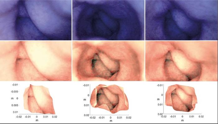
Researchers at the Norwegian University of Science and Technology (NTNU) have developed a groundbreaking technique to create 3D models of the colon from a single image. This innovation could revolutionize how doctors detect and diagnose gastrointestinal diseases, potentially offering a less invasive alternative to traditional colonoscopies.
The study, recently published in the Journal of Imaging, demonstrates the ability to reconstruct an accurate three-dimensional model of a patient’s colon using just one image from a capsule endoscopy camera. This technology could significantly improve the detection of abnormalities in the gastrointestinal tract, leading to faster diagnoses and more effective treatment of conditions like bowel cancer.
From 2D to 3D: A Technological Leap
The research team, led by Pål Anders Floor from NTNU’s Department of Computer Science, used an artificial colon, endoscopic images, and a specialized algorithm to create their 3D models. The key to their success lies in a mathematical model called the shape-from-shading algorithm (SFS), which can reconstruct a three-dimensional shape from a single, two-dimensional image.
“We show that by carefully calibrating and pre-processing the images, we can obtain a good 3D model based on just one image – even if the image is full of noise and visual distortion,” Floor explained.
This breakthrough is particularly significant given the limitations of current capsule endoscopy technology. While capsule cameras have been in use for over two decades, their effectiveness has been hampered by low-quality images and image noise. The NTNU team’s work could breathe new life into this smart technology.
Improving Patient Care and Diagnosis
The potential benefits of this technology for both doctors and patients are substantial. The 3D models allow specialists to examine the digestive tract from various angles on a computer, enabling better planning and more precise interventions. Doctors can even practice difficult procedures beforehand, reducing the risk of complications.
“The 3D model can be used to adjust the lighting in an original image so that darker areas become better illuminated and more detail can be seen,” Floor noted. This enhanced visibility could lead to more accurate diagnoses and earlier detection of diseases like bowel cancer, which is the second most common type of cancer in Norway.
While it’s too early to declare this a definitive breakthrough in the fight against bowel cancer, the potential is clear. Floor and his colleagues are continuing to refine their methods and conduct further research to achieve statistically valid results.
The technology’s applications extend beyond gastroenterology. The researchers believe their technique could be valuable in fields such as cultural heritage preservation, robotics, and other areas of medical diagnostics where generating 3D models from images would be beneficial.
As this technology continues to develop, it offers hope for less invasive, more accurate diagnostic tools in gastrointestinal medicine. For the millions of people who dread traditional colonoscopies, these 3D colon models could represent a welcome alternative, potentially increasing early detection rates and improving outcomes for patients with bowel diseases.


