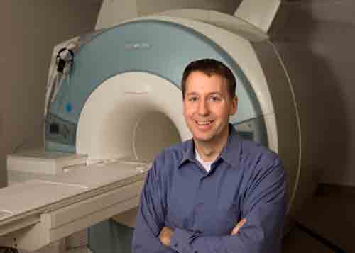
A joint BYU-Utah research team is developing a new breast cancer screening technique that has the potential to reduce false positives, and, possibly, minimize the need for invasive biopsies.
Led by BYU electrical engineer Neal Bangerter and University of Utah collaborators Rock Hadley and Joshua Kaggie, the group has created an MRI device that could improve both the process and accuracy of breast cancer screening by scanning for sodium levels in the breast.
“The images we’re obtaining show a substantial improvement over anything that we’ve seen using this particular MRI technique for breast cancer imaging,” said Bangerter, senior author on a study detailing the method in academic journal Magnetic Resonance in Medicine.
Specifically, the device is producing as much as five-times more accurate images than previous efforts with an emerging methodology called sodium MRI.
Currently, there are two clinical imaging methods widely used for screening breast cancer: mammograms and proton MRI scans.
X-ray mammography is the most common screening tool, but the procedure involves x-ray exposure and is generally unpleasant. Mammograms are relatively inexpensive, but they still lead to biopsies when something suspicious is detected.
Because of their increased sensitivity, proton MRI scans are generally used to further examine suspicious areas found by mammograms. However, they can produce false positives leading to unnecessary interventions.
Sodium MRI has the potential to improve assessment of breast lesions because sodium concentrations are thought to increase in malignant tumors. Bangerter and his team believe that the addition of sodium MRI to a breast cancer screening exam could provide important additional diagnostic information that will cut down on false positives.
The team has developed a new device used for sodium imaging that is picking up a level of detail and structure not previously achieved.
“This development by Dr. Bangerter and his group represents a major advance in the field of multinuclear MRI of the breast,” said Stanford Professor of Radiology Bruce Daniel. “He and his group have invented a way to dramatically boost the sodium signal from the breast, enabling much better, higher resolution sodium MR images to be obtained. This should open the door to new avenues of research into breast cancer.”
So far, the novel technique returns high-quality images in only 20 minutes, improving the odds that sodium MRI breast scans could be implemented clinically.
The MRI team’s goal is to produce a device capable of obtaining both excellent sodium and good proton images without requiring the patient being screened to be repositioned for multiple scans.
“This method is giving us new physiological information we can’t see from other types of images,” Bangerter said. “We believe this can aid in early breast cancer detection and characterization while also improving cancer treatment and monitoring.”
Bangerter’s major University of Utah collaborators include Hadley, Glen Morrell and Dennis Parker, all professors of radiology at the University of Utah’s Center for Advanced Imaging Research, and Kaggie, a graduate research assistant who is the primary author on the recent academic journal article detailing the work.


