Scientists have developed a powerful new technique that could transform our understanding of the brain’s intricate wiring system. The breakthrough method, called Light-microscopy-based Connectomics (LICONN), enables researchers to map the brain’s complex neural networks at the nanoscale while simultaneously identifying specific molecules within those connections. This innovative approach, detailed in a new study published in Nature, brings scientists one step closer to comprehensively understanding how brain cells communicate and function together.
Bridging the Gap Between Structure and Function
Until now, researchers studying brain connectivity faced a challenging trade-off: they could either see the detailed physical structure of neural connections using electron microscopy (EM) or identify specific molecules using light microscopy—but not both simultaneously at the resolution needed to trace individual connections.
LICONN overcomes this limitation by combining innovative tissue preparation with artificial intelligence. The technique expands brain tissue samples approximately 16-fold while preserving their structural integrity, then uses machine learning to analyze the expanded samples and reconstruct their intricate cellular architecture.
“The brain is made up of an incredibly dense, complex and fine-grained arrangement of neurons with support cells, which together constitute a functional network that enables brain function,” the researchers explain in their study. This new approach allows scientists to visualize this arrangement with unprecedented clarity and molecular context.
How LICONN Works
The technique builds on a method called expansion microscopy, which physically enlarges tissue samples to reveal details too small to see with conventional microscopes. The LICONN approach significantly refines this process through a series of chemical innovations:
- Brain tissue is embedded in a specially engineered expandable hydrogel that preserves cellular structures
- The tissue-hydrogel hybrid is expanded approximately 16-fold, bringing nanoscale features into view
- Standard fluorescence microscopy captures the enlarged structures at high resolution
- Deep learning algorithms reconstruct the neural networks with 92.8% accuracy
- Molecular tags identify specific proteins at synapses and other cellular structures
This combination allows researchers to visualize details as small as 20 nanometers using standard microscopes—a resolution previously achievable only with much more complex and expensive specialized equipment.
Seeing Brain Connections in a New Light
Traditional approaches for mapping brain circuits with electron microscopy have made enormous progress in recent years but have important limitations, particularly in extracting molecular information. By contrast, LICONN directly visualizes both structural connections and molecular details in the same sample.
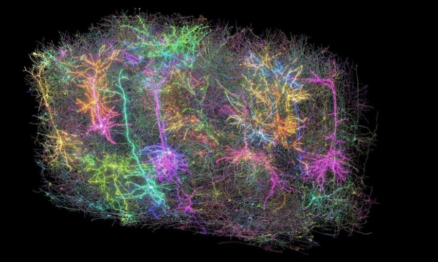
The researchers demonstrated LICONN’s capabilities by mapping a volume of brain tissue approximately 1 million cubic micrometers in size from the mouse cerebral cortex. Within this volume, they identified individual neurons, their connections, and the molecular makeup of those connections.
The technology revealed stunning details of brain architecture, including the precise arrangement of synaptic proteins spaced less than 100 nanometers apart—about 1/1000th the width of a human hair. It also identified periodic patterns of proteins along axons, the long projections that neurons use to send signals, spaced at intervals of around 89 nanometers.
Making Advanced Brain Mapping More Accessible
Perhaps most significantly, LICONN makes high-resolution brain mapping accessible to more researchers. While electron microscopy requires specialized equipment and expertise, LICONN works with standard laboratory microscopes and techniques.
“LICONN is highly accessible,” the researchers note. “Acquisition is driven by broadly available conventional light-microscopy hardware… and, although LICONN sample preparation introduces new strategies to achieve high-fidelity tissue expansion, the protocol is not fundamentally more complex than previous expansion techniques that have been widely adopted.”
This accessibility could dramatically accelerate research into brain structure and function across the neuroscience community.
Beyond Basic Connections
The molecular information provided by LICONN opens new research avenues beyond mapping basic connections. In their study, the researchers demonstrated several applications, including:
Identifying excitatory versus inhibitory synapses based on their molecular composition, providing insight into the balance of neural activation and suppression. The team found that approximately 90% of inputs to dendrites (branched extensions of nerve cells) were excitatory, while 10% were inhibitory—consistent with previous observations.
Visualizing electrical connections called gap junctions, which are difficult to identify with electron microscopy but are crucial for understanding how groups of neurons synchronize their activity.
Examining specialized cellular structures like primary cilia—antenna-like projections that cells use for signaling—and comparing their characteristics between normal mice and those with a genetic mutation linked to epilepsy and intellectual disability.
Looking to the Future of Brain Mapping
While the current implementation of LICONN has mapped volumes similar in size to those studied with electron microscopy, the researchers envision scaling the technology to map much larger brain regions. They suggest that combining LICONN with techniques to measure gene expression directly in tissue could provide an even more comprehensive picture of brain function.
“By integrating connectivity with in situ molecular information from individual cells, LICONN presents a viable path towards multimodal descriptions of mammalian brain cells, including morphology, connectivity (including electrical connections), physiology and gene expression,” the researchers conclude.
As neuroscientists continue to unravel the extraordinarily complex wiring of the brain, technologies like LICONN that bridge the gap between physical connections and molecular function promise to accelerate our understanding of how the brain works—and potentially what goes wrong in neurological disorders.
Discover more from NeuroEdge
Subscribe to get the latest posts sent to your email.