

The dromedary camel has spent millions of years being battered by bacteria, viruses and parasites across some of the harshest terrain on Earth. It rarely loses. While sheep and goats in the same Omani pastures fall to foot-and-mouth disease, tetanus and other common infections, camels shrug them off with an equanimity that has long puzzled immunologists. Unique single-domain antibodies — nanobodies, discovered in the late 1980s — explained part of this resilience. But researchers at Sultan Qaboos University now think there’s more to the story, buried in the camel’s genome, waiting to be turned into medicines.
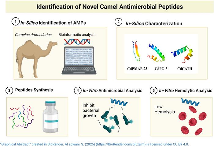
Their new paper, published in Frontiers in Immunology, reports the identification of three peptides from dromedary camel DNA that can tear apart the membranes of multidrug-resistant bacteria, including MRSA and a strain of E. coli resistant to multiple classes of antibiotics. The findings are preliminary — no animal trials yet, no clinical applications imminent — but they add another chapter to a growing body of evidence that the natural world still holds reservoirs of antimicrobial chemistry we haven’t touched.
The timing matters. The antibiotic pipeline has been running dry for decades. Most conventional drugs work by binding to specific molecular targets inside bacteria — interfering with cell-wall synthesis, jamming ribosomes, blocking DNA replication. That precision is also a vulnerability: a single mutation in the target site can render an antibiotic useless. Bacteria are extraordinarily good at acquiring such mutations, sharing resistance genes across species, evolving around whatever chemistry we throw at them. The World Health Organisation has been warning of a post-antibiotic era for years. The warnings are no longer speculative.
Antimicrobial peptides offer a different kind of attack. Rather than sneaking into bacterial cells and interfering with their machinery, these short protein chains simply destroy the cell membrane itself — electrostatically attracted to the negatively charged surface of bacterial cells, they fold into helical shapes on contact, wedge into the lipid bilayer, and punch holes in it. Bacteria can’t easily develop resistance to this kind of assault; there’s no single binding site to mutate, no one molecular lock to change.
Cathelicidins are the best-studied class of these peptides in mammals. The human version, LL-37, is found in skin cells, neutrophils, and the lining of the lungs and gut. It’s been known since the 1990s that cathelicidins turn up in cattle, horses, pigs and chickens — each species producing its own structural variants, tuned to local microbial threats. What hadn’t been systematically explored was whether dromedary camels had their own. Given the animals’ remarkable infection resistance, that seemed like an oversight worth correcting.
The Sultan Qaboos team — working with collaborators at the Karolinska Institutet in Stockholm — started in silico, mining the camel genome for sequences resembling known cathelicidins from other Camelidae: Bactrian camels, wild camels, and alpacas. Three candidates emerged. They named them CdPMAP-23, CdPG-3, and CdCATH — the Cd prefix marking their camel origin.
All three share the structural hallmarks of cathelicidins: a conserved “cathelin-like domain” at one end, packed with four cysteine residues, and a variable antimicrobial region at the other. This architecture is ancient and conserved across mammals. The conserved end appears to be involved in regulating when and where the peptide gets activated; the variable end is where the actual bactericidal action happens, which may be why it varies so much between species.
The structural differences between the three camel peptides turn out to matter enormously for what they do. CdPG-3 and CdCATH both fold into amphipathic alpha-helices when they encounter bacterial membranes — one face hydrophobic, the other charged, allowing them to insert into and destabilise the lipid bilayer. CdPMAP-23 doesn’t do this; it stays in a disordered “random coil” conformation even in membrane-like conditions, which probably explains why it performed more modestly in the subsequent experiments. Circular dichroism spectroscopy — a technique that measures how proteins rotate polarised light, revealing their secondary structure — confirmed this. In the presence of SDS (a membrane-mimicking detergent) and bacterial lipopolysaccharide, CdPG-3 adopted an alpha-helical structure roughly 78 per cent of the time. CdCATH reached around 56 per cent helicity under the same conditions. CdPMAP-23 barely budged.
The antibacterial assays bore this out. Against six bacterial strains — including standard reference cultures of Staphylococcus aureus, E. coli and Klebsiella pneumoniae, plus drug-resistant MRSA and multidrug-resistant E. coli — CdPG-3 and CdCATH both performed impressively. Against the carbapenem-resistant K. pneumoniae strain ATCC 1705, which produces an enzyme called KPC that chews up an entire class of last-resort antibiotics, CdCATH achieved complete suppression of bacterial growth at 40 micromolar concentration. CdPG-3 reached a 3.5 log-fold reduction at 160 micromolar. These are not trivial findings.
The electron microscopy images are, frankly, striking. Untreated E. coli cells look exactly as you’d expect — smooth, regular, intact. After 30 minutes of CdCATH treatment, most cells are barely recognisable. Membranes have ruptured. Internal structures have spilled out. What remains looks like the aftermath of something explosive. CdPG-3 does similar damage, producing pores and surface blebbing. Even CdPMAP-23, the weakest performer in the group, left cells visibly swollen and morphologically abnormal.
The safety question — how selectively these peptides attack bacteria rather than host cells — was addressed through hemolysis assays, testing whether the peptides destroy red blood cells from humans, camels, goats and chickens. The results are mixed, and they’re candid about it. At therapeutic concentrations (below 20 micromolar), CdPG-3 showed minimal hemolytic activity in human blood, staying well under the 5 per cent threshold considered clinically acceptable. CdPMAP-23 was also clean. CdCATH is more problematic: above 20 micromolar in human cells, hemolysis increases substantially, exceeding 50 per cent at 80 micromolar. That’s a concern that would need to be engineered away before any clinical use. Interestingly, all three peptides were completely non-hemolytic against camel red blood cells at all concentrations tested — a reflection, apparently, of the unusual lipid composition of dromedary erythrocytes, which contain high cholesterol levels that make the membrane unusually resistant to disruption.
The paper is careful about its limitations. Only six bacterial strains were tested. Antifungal and antiviral activity, suggested by computational predictions, wasn’t experimentally verified. The immunomodulatory properties of cathelicidins — their ability to recruit immune cells, promote wound healing, and modulate inflammation — were similarly left for future work. These aren’t minor gaps; cathelicidins in other mammals do as much through immune signalling as through direct bacterial killing, and understanding the full picture matters for any drug development effort.
What the study does establish, cleanly, is that dromedary camels produce cathelicidin-type antimicrobial peptides that are real, active, and structurally capable of the membrane disruption mechanism their sequence analysis predicted. That’s the foundation from which medicinal chemistry can work: modifying the amino acid sequences to reduce hemolysis, increase stability in the bloodstream, and optimise potency against specific resistant pathogens.
The camel, in other words, has been doing its own antibiotic research for a very long time. We’re only just starting to look at the results.
Study link: https://www.frontiersin.org/journals/immunology/articles/10.3389/fimmu.2026.1745714/full