

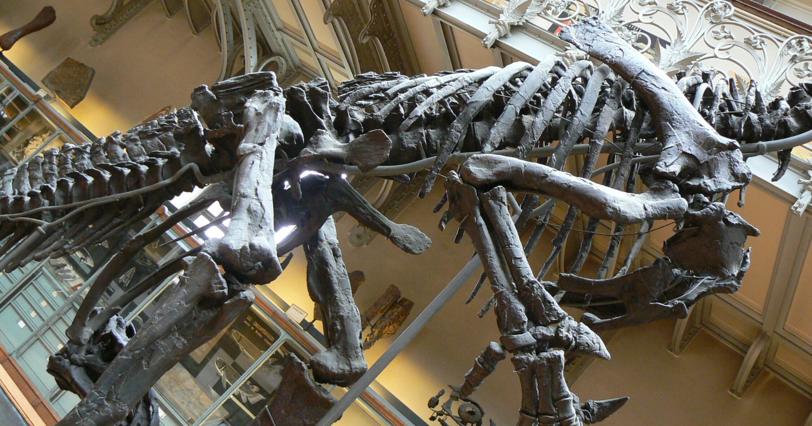
Six researchers in Spain have described a swollen patch on the shin bone of an Iguanodon bernissartensis and concluded that the bone was reacting to an ulcer in the skin above it. They report it, to their knowledge, as the first case of that kind of infectious damage identified in a dinosaur, in a paper published in Royal Society Open Science on 26 August.
The paper’s own verb for the diagnosis is careful. The lesion, it says, is consistent with a periosteal reaction and cortical thickening caused by a skin ulceration that reached the bone. It gets there by elimination. The authors open with several possible causes, among them a fracture callus, bone bruising, a tumour and a traumatic infection, and work the first two out of the running, ruling out several named tumours one at a time. The fourth is the one the conclusion keeps, calling the aetiology consistent with a traumatic-infectious episode.
The paper does not agree with itself about the swelling
The specimen, catalogued CMP-11/8, is a complete right tibia with a preserved length of 1,023 millimetres. The shaft is straight and the astragalus is fused to the distal end.
The abnormal growth sits in the lower half of the bone. It measures 174 millimetres long and 117 millimetres wide, with a maximum thickness of 17.3 millimetres, and the authors put it at roughly 17 percent of the tibia’s total length. Viewed from the side, it projects 21.9 millimetres beyond the rear margin of the bone.
Its position is where the paper stops adding up. A single sentence puts the start of the growth 109 millimetres under the tibia’s mid-length and has it extending up to 446 millimetres from the distal end; the sentence after that gives its length as 174 millimetres. Mid-length on a 1,023 millimetre bone falls 511.5 millimetres from the distal end, so 109 below that is 402.5, and the gap between 402.5 and 446 is 43.5 millimetres, not 174. That arithmetic is ours, and the three figures cannot all be right as printed. The 174 millimetre length is the one that closes, since 174 of 1,023 is 17.0 percent.
The surface is smooth and swollen rather than broken, with what the paper calls a well-defined lobulated morphology, thicker at the bottom and tapering as it runs up the bone, and the shaft is neither shortened nor deformed. Where the growth actually lies is a second disagreement, two statements against one. The abstract puts it on part of the tibia’s posteromedial side and the results section gives it most of the inner face plus the entire back, while the later argument against a fracture calls it exclusively medial with no trace on the outer side.
The bone came from site 11 of the Mas de la Parreta quarry at Morella, in Castellón province, out of blue-grey sandy siltstones in the upper Barremian Arcillas de Morella Formation, alongside at least two partially disarticulated I. bernissartensis carcasses, theropod and crocodile-relative teeth, and turtle shell.
George Albert Boulenger named the species in 1881 on material from a Belgian coal mine at Bernissart, where thirty relatively complete skeletons were found 322 metres underground, the bones still in their original position. Around Morella, Iguanodon remains were first reported in 1873, this species was reliably identified only in 1982, and about ten partial skeletons have since come out of the formation. Diseased bone stays scarce: before this tibia, one pathological vertebra from the same quarry had been reported, and only preliminarily.
A hospital scanner stood in for the saw
Cutting a thin section is, in the paper’s words, a valuable technique for reading what happened inside a fossil bone, and the team ruled it out because it means destroying part of the specimen. They took the tibia to the radiology department of the Hospital General Universitari de Castelló instead and put it through a medical Canon CT scanner at 130 milliamps and a peak voltage of 100 kilovolts, at 0.5 millimetre voxels.
The clinical setting is not incidental. Four of the six authors, the lead author Javier Salas-Herrera among them, are at the evolutionary biology group of Spain’s national distance university, UNED. The other two are hospital clinicians: Santiago F. Marco Domenech in radiodiagnosis and Ricardo Tosca-Segura in paediatrics.
The scan showed that the parts of the shaft outside the growth are well preserved, with cortical and spongy bone still distinguishable. Inside the growth, the cortex shows low thickening and moderate expansion, slightly deforming the inner surface beneath it. The lesion stops short of the marrow cavity, and the cortical bone under the periosteum shows hyperdense patches in both sagittal and axial views.
The substitution has a cost the authors do not dwell on. Because histology was declined, everything downstream rests on gross shape and CT density at half-millimetre voxels rather than on bone microstructure, which is what would show whether the bone was laid down quickly or slowly and whether the reaction was still active when the animal died.
What the scan settled, and what it left open
A healing fracture was the first candidate, and one reported many times over in other dinosaur long bones. It is also the candidate the paper excludes most firmly. No clear disruption of the cortex or the inner surface is recognised, of the kind that marks a bone repairing itself, and a full callus should thicken the entire width of the shaft, whereas the argument here treats the growth as exclusively medial with the outer side clean. That is the description the paper contradicts in its own results section, and the exclusion depends on it.
A healed stress fracture, a routine finding in the shins of runners and children, can mimic the same smooth thickening; it is ruled out because the scan shows none of the linear radiolucent areas or the associated inner thickening that go with one. A greenstick fracture, diagnosed before in juvenile dinosaur bones, gets no verdict of its own, the paper noting only that this tibia is plainly an adult’s. Bone bruises are precluded on position: they commonly develop close to joint surfaces, and this lesion is mid-shaft, with the inner bone surface running continuously beneath it.
Tumours take longest. The absence of the aggressive periosteal reaction typical of malignancy, plus the well-circumscribed location of the lesion, excludes periosteal osteogenic osteosarcoma, Ewing’s sarcoma and Langerhans cell histiocytosis, and the paper’s list of tumours is explicitly open rather than exhaustive. Osteoid osteoma, a benign growth built around a small radiolucent core, is ruled out because the scan reveals no such core. Osteofibrous dysplasia, which produces a soap-bubble pattern of cavities and can bow the bone, is ruled out because this tibia shows neither.
Three candidates are handled more softly, and the difference matters. Osteoblastoma, the larger variant of osteoid osteoma, gets only the observation that it tends to appear in vertebrae rather than long bones. Chronic osteomyelitis, an infection beginning in the bone, sometimes lays down a dense sheath called an involucrum, and the scan found no such pattern, but the paper records that negative and stops; it does not say osteomyelitis is excluded. And traumatic infection, the fourth of the causes the paper opens with, never gets a verdict of its own at all, because it is the reading the conclusion goes on to adopt.
The surviving diagnosis rests on shape and density
What is left is a lesion sitting just outside the cortex, with a periosteal reaction running parallel to the length of the bone. The authors write that it certainly resembles a primary periostitis and exhibits some features indicative of an advanced infectious process that began in soft tissue and worked inwards. They also concede, in the same passage, that several aetiologies can be attributed to the bone characteristics of a periostitis, which is the paper saying its central finding is not specific to any one cause.
The comparison they reach for is clinical. Continuous reactive bone plus a thickened, hardened cortex resembles an osteoma-like lesion, which is in turn comparable to the bone changes recorded beneath tropical skin ulcers with a clinical history of a year or less. Those ulcers usually affect the middle third of the tibia and fibula; the paper places this lesion at the midshaft, in the bone’s distal half. Pollen and freshwater algae from the deposit point to a warm, humid subtropical to warm-temperate climate of the kind such ulcers favour.
What the paper does not have is a microbe. It reports no attempt to recover, culture or sequence microbial material from the bone, and presents no such evidence. The polymicrobial part of the label is borrowed from living patients, in whom fusiform bacilli and spirochetes are often isolated from tropical ulcers, and the aside that reptiles today can carry spirochetes such as Leptospira and Borrelia is an argument for plausibility, not a finding about this animal.
The paper is titled “First occurrence of polymicrobial ulcer in dinosaurs”. Its conclusion calls the lesion the result of a skin ulceration, “potentially a polymicrobial ulcer (tropical ulcer)”. Those are not the same claim, and the conclusion is the one the evidence reaches.
Timing is unsettled in the same way. The abstract describes an ulceration “in an advanced stage” and the discussion an “advanced infectious process”, while the conclusion twice calls the episode acute, and the paper does not reconcile the two. How the sore began drifts as well: the discussion has the infection originating in soft tissue, and the conclusion calls the aetiology consistent with a traumatic-infectious episode, which is a different account of the first event.
The claim of a first is a claim about the published record. It is hedged “to our knowledge” three times, in the abstract, the introduction and the conclusion, and scoped four different ways in those same four places, from “polymicrobial ulcer” in the title to “this type of pathological response to a bacterial infection” in the abstract. What the authors say they established is that nobody they could find has previously reported the bone signature of a dinosaur skin ulcer, which is not a claim that such ulcers were rare.
Two smaller loose ends are worth knowing about. The scans are described as available from a Spanish repository on request, while the reference given for them is a Dryad deposit whose DOI returns an error and which Dryad’s own records show as never published. And the paper gives no absolute age for the specimen or the formation, figure captions included: the bone is placed in the upper Barremian and nothing more, so any figure in millions of years attached to this story has come from outside it.
A limp that cannot be measured
The authors offer one line about the animal itself, as a supposition. It is reasonable, they write, to suppose that an ulcer eating down to the periosteum may have caused persistent pain, aggravated by walking and by muscle pressure across the site, and that scarring over such an area would have left the leg working less well than it should.
Nothing in a single tibia tests that. A thin section would separate a fast reaction from a slow one and an active lesion from a healed one, at the price of a slice through the growth. Failing that, the roughly ten partial skeletons already out of the Morella formation, and whatever the quarry yields next, are where a second case would have to come from.