

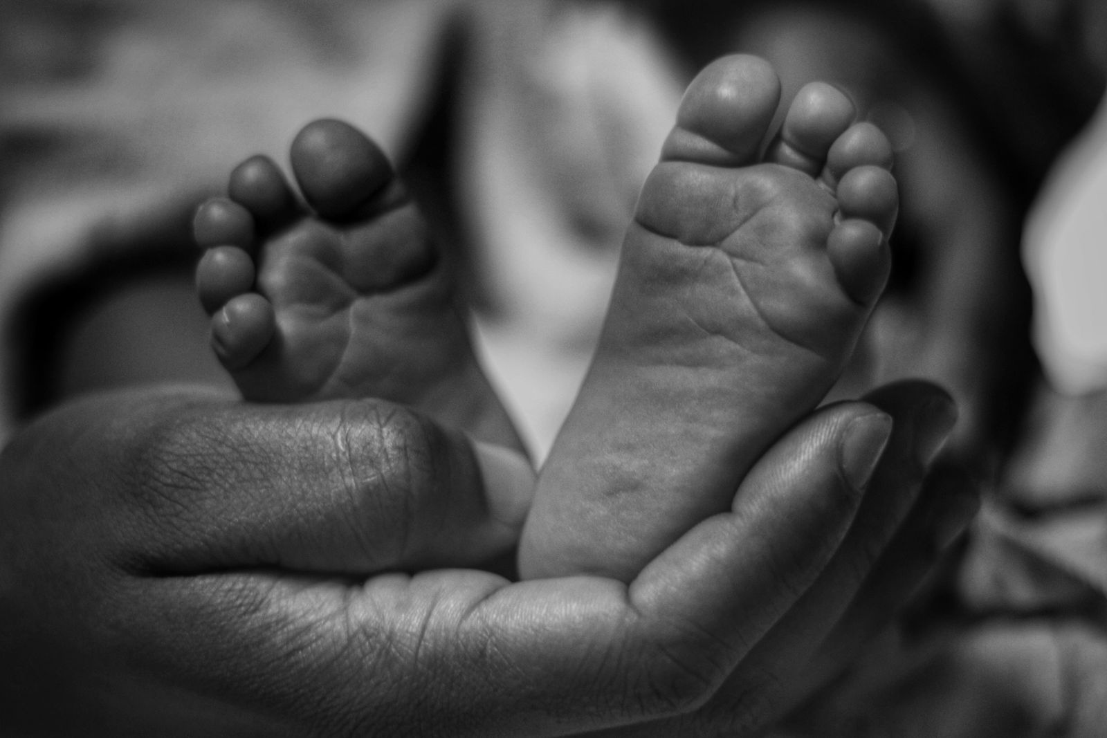
A newborn placed on a scale weighs less than a Thanksgiving turkey and carries inside that small body roughly 300 separate bones. An adult skeleton, by contrast, contains 206. The missing hundred or so did not vanish. They fused. The skull plates that overlapped as the baby squeezed through the birth canal, the spinal segments that let a six-month-old arch and roll, the three pelvic bones that met at the hip — all of them slowly knit together into the single structures an adult now takes for granted.
The body you sit in was assembled from parts that once moved independently while you learned to hold up your own head.

The skull arrives in pieces
The clearest example is the top of the head. A newborn’s cranium is not one dome but a set of curved plates separated by ribbons of fibrous tissue called sutures, and by wider gaps called fontanelles — the soft spots parents are warned about. Those spaces exist because the human brain is going to do something extraordinary in the months after birth.
At delivery, the brain is roughly a quarter of its eventual adult size. It doubles within the first year. By age five it has reached about 90 percent of its final volume. A rigid skull could not accommodate that expansion. So the skull arrives unfinished.
Five major sutures divide the infant cranium: the coronal, running ear to ear; the sagittal, tracing the crown from front to back; the lambdoid at the rear; the metopic between the frontal bones; and the squamous above each ear. During birth, these seams let the plates slide and even overlap, narrowing the head enough to pass the pelvic outlet. In the following months, they act as expansion joints for the growing brain.
The metopic suture usually closes first, sometime before a child’s second birthday. The anterior fontanelle — the diamond-shaped soft spot near the crown — typically shuts between 12 and 18 months. The others fuse gradually across childhood and adolescence. By adulthood, the 22 bones of the skull are locked together so tightly that the seams look like cracks in porcelain.
Why the count drops from 300 to 206
The skull is only one theater of this quiet consolidation. A newborn’s spine contains separate segments in the sacrum and coccyx that will later fuse into two solid structures. The five sacral vertebrae, distinct at birth, merge into a single triangular bone at the base of the spine by the late teens to mid-twenties. The four (sometimes three, sometimes five) tiny coccygeal segments fuse into the tailbone.
The hip tells the same story. Each side of the adult pelvis — the innominate bone — is actually three childhood bones: the ilium, the ischium, and the pubis. In an infant they meet at the hip socket in a Y-shaped strip of cartilage called the triradiate cartilage. That cartilage does not ossify into solid bone until the mid-to-late teenage years. Long before then, it has been quietly widening the acetabulum to hold a growing femoral head.
Long bones do their own arithmetic. The femur, the humerus, the tibia — each begins as a shaft with separate caps at either end, called epiphyses, joined to the main shaft by a plate of growing cartilage. Those growth plates are why a broken arm at age nine heals differently from a broken arm at 40, and why a pediatric radiologist can read a hand X-ray and estimate a child’s age within months. The plates close in a predictable sequence through the teens and early 20s, converting what were three or more separate ossification centers into one adult bone.
Add all of these mergers together and the arithmetic works out. The count of about 300 at birth is not a single agreed-upon number — anatomists differ on how to tally cartilage that is on its way to becoming bone — but the drop to 206 by adulthood is standard.
Cartilage is the scaffolding
The material holding the infant skeleton together is mostly cartilage — the same rubbery tissue that caps your knees and shapes the tip of your nose. Cartilage is flexible, avascular, and grows readily. Bone is rigid, vascular, and grows only at its edges. The developmental trick of the mammalian skeleton is to lay down cartilage first, in roughly the right shape, and then slowly replace it with bone through a process called endochondral ossification.
This is why a baby feels boneless in places an adult does not. The wrist of a one-year-old contains almost no bone visible on X-ray, because the eight carpal bones are still cartilaginous nubs. They calcify one by one across childhood, in an order so reliable that radiologists use it as a clock.
The ribs, too, retain cartilage where they meet the sternum, which is why an adult chest can still flex with each breath. Some of that infant flexibility never fully goes away. It just narrows to specific joints.
An evolutionary compromise
Why be born so unfinished? The answer sits at the intersection of two pressures that shaped the human lineage. Humans have unusually large brains and, thanks to bipedal walking, unusually narrow pelvises. A fully developed head cannot pass through a fully developed birth canal. Something has to give.
What gives is the timing of skull ossification. Human infants are born neurologically helpless — they cannot walk, cannot hold their heads up, cannot regulate their own temperature well — but their skulls are still soft enough to deform during birth and then keep expanding afterward. A chimpanzee newborn, by comparison, arrives more neurologically mature but with a brain that will grow proportionally less.
The human skull has been reshaping itself across millions of years of evolution. Since the split from the chimpanzee lineage roughly 6 to 7 million years ago, the braincase has ballooned, the face has retreated beneath it, and the ridges that once anchored heavy chewing muscles have smoothed away. According to research on human facial evolution, the modern human face is significantly reduced compared to great apes, giving the human skull its distinctive globular shape. The globular shape of the adult human skull is a signature of that history — and it is a shape only achievable if the skull arrives in fragments and finishes itself outside the womb.
Neanderthal babies grew the same way
The closest test of what makes human infant development distinct comes from the fossils of a cousin species. Neanderthals shared this basic architecture — separate bones fusing over time — and recent analyses suggest their earliest development tracked ours closely.
A set of Neanderthal infant remains from Sesselfelsgrotte, a rock shelter in Bavaria, has given researchers a rare window into the first months of Neanderthal life. The fossils date to roughly 50,000 to 75,000 years ago, and micro-CT scans of the bones and teeth have let researchers reconstruct fetal and infant growth in unusual detail. As Earth.com reported, the growth patterns of these Neanderthal infants overlap substantially with those of modern human babies, suggesting the pace of early skeletal development was broadly shared between the two species.
A parallel analysis, also published by Discover, traced fetal growth in the same Bavarian fossils and identified possible signs of metabolic stress recorded in the enamel of infant teeth — some of the earliest such evidence in the hominin record. The finding sharpens a picture in which Neanderthal newborns, like ours, arrived with unfused skulls and cartilaginous joints that would consolidate over years.
That the two species developed on such similar schedules matters, because the two lineages overlapped in space and time. Modeling of radiocarbon-dated artifacts from 17 sites across France and northern Spain estimated that Homo sapiens and Neanderthals co-existed in the region for between 1,400 and 2,900 years before Neanderthals disappeared roughly 40,000 years ago. For most of that overlap, infants of both species were being born with the same soft-plated skulls, learning to sit up as their sutures narrowed.
What fuses, and when
A rough timetable of consolidation gives a sense of how long the process takes.
The metopic suture in the forehead closes between about nine months and two years. The anterior fontanelle shuts by roughly 12 to 18 months. The posterior fontanelle at the back of the head closes earlier, often by two or three months of age. The sacrum fuses across the late teens and 20s. The triradiate cartilage of the pelvis ossifies around ages 11 to 15. The medial end of the clavicle, the last major growth plate to close, does not fully finish until the late 20s.
That final detail has a forensic use. Because the medial clavicle closes so late and so predictably, forensic anthropologists examining unidentified remains often use it to distinguish a 22-year-old from a 30-year-old. The bone that was still knitting itself together when you finished graduate school is, in effect, a birth certificate.
The pieces you carry
Run a finger along the top of your own skull and you can sometimes feel a faint ridge where the parietal bones met the frontal — the ghost of the coronal suture. Press on the base of your spine and you are touching the fused remnant of five vertebrae that were once distinct. The socket where your femur pivots was carved out of three separate bones cooperating in cartilage.
None of this is metaphor. The adult human skeleton is a mosaic that finished assembling itself only in the last decade or so of adolescence, and in a few places even later. The skull plates that let you be born have long since locked; the pelvic bones that shifted as you learned to walk have long since fused; the growth plates that added an inch a year during middle school have sealed shut. What remains is a single, articulated frame — a fossil of the child who once contained a hundred more moving parts than the adult it grew into.