

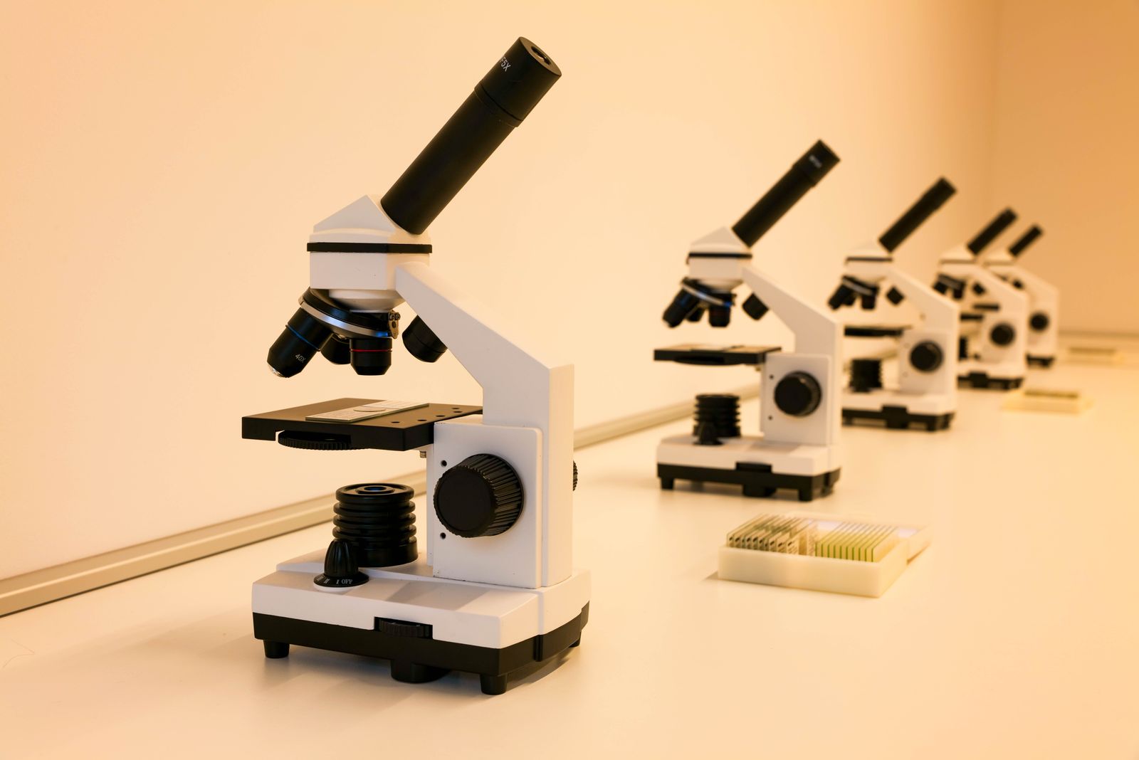
In 1665, an English polymath named Robert Hooke placed a paper-thin shaving of cork under a brass-and-leather microscope he had built himself, held a candle behind a glass globe of water to focus the light, and saw what no one had ever described before: thousands of tiny, empty, box-like compartments packed together like a honeycomb. He wrote that they reminded him of the small bare rooms in a monastery. He called them cells. The word has stuck for more than three and a half centuries.
The drawing and description appeared in Micrographia, published in 1665, a folio-sized book of copperplate engravings depicting fleas the size of dinner plates, the compound eyes of flies, the point of a needle, and the surface of a piece of cork. It was the first bestseller of the scientific revolution.

A carpenter’s son with a talent for lenses
Hooke was a young man when the book came out. He was the curator of experiments at the Royal Society, which meant that every week he had to invent, build, and demonstrate something new for the fellows to gawp at. A microscope was one of his tools of the trade.
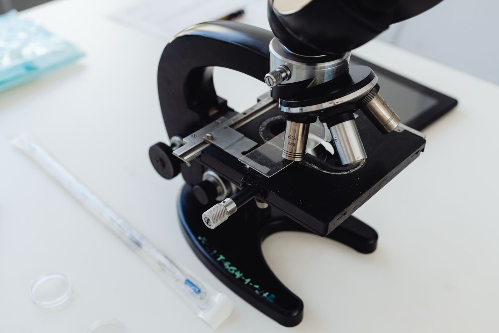
The instrument he used for the cork observation was a compound microscope, with a tube of pasteboard covered in leather and gilded, two lenses inside, and a separate lamp system he had engineered himself. Because early lenses smeared the image with colour fringes and blur, Hooke devised a workaround: a brine-filled glass sphere that concentrated candlelight into a bright, cold beam on the specimen. The Atlantic has described his rig as a piece of hacked optical plumbing, closer to a jeweller’s workbench than a laboratory instrument.
His best magnification was somewhere around 50x. Modern super-resolution microscopes resolve far smaller features. But 50x was enough to see the ghost of a plant’s architecture.
Why the cork looked like a monastery
Cork is the outer bark of the cork oak, Quercus suber. By the time it is stripped from the tree, the living contents of its cells have long since died and drained away, leaving only the boxy walls of dead plant tissue — essentially a dried scaffold of lignin and suberin. That is why Hooke saw empty rooms rather than the messy, fluid-filled interior of a living cell.
He counted the compartments. In one square inch, he estimated, there were a great number of them. He wrote that the little boxes reminded him of the cells — the small sleeping chambers — that monks occupied in a monastery. The Latin root, cella, means a small room or storeroom, and it had already been used for centuries to describe honeycomb chambers and prison cells. Hooke simply extended the metaphor down a scale nobody had previously visited.
He did not realise he was looking at the fundamental unit of life. He thought he had found a clever architectural trick of plant tissue that explained why cork was so light and buoyant. The idea that every living thing — oak, elephant, human, bacterium — is built out of these compartments would not arrive until decades later.
The book that made London stare
Micrographia was not a dry scientific tract. It was a spectacle. The engravings folded out to more than a foot across. A flea, drawn from Hooke’s own observations, sprawled across the page in armoured, spiky detail that horrified readers who had lived their whole lives with the insect in their beds and never seen its face. A louse gripped a human hair thicker than its own body. The compound eye of a grey drone-fly resolved into a lattice of thousands of hexagonal facets.
The diarist Samuel Pepys, then a rising naval administrator in his early thirties, bought a copy and later described it as the most ingenious book that I ever read in my life — a rare superlative from a man who spent his evenings at the theatre, at coffeehouses, and in candlelit rooms full of borrowed books. Pepys’s copy is often held up as evidence of how quickly Hooke’s images crossed from the Royal Society’s fellows into the reading rooms of London’s professional class.
Pepys’s world in 1665 was about to be turned inside out. As the year wore on, he was writing uneasy entries about the plague creeping through the city. His diary records great fears about the sickness in the city. By late summer the death toll had reached thousands of people a week.
A flea on the page and a flea in the streets
One of the strangest coincidences of 17th-century science is that the most famous engraving in Micrographia — Hooke’s monumental fold-out flea, all glossy carapace and jointed legs — was published the same year the rat flea, Xenopsylla cheopis, was silently seeding the Great Plague of London. Nobody in 1665 knew that fleas carried Yersinia pestis. That connection would not be established until the late 19th century.
Pepys worried about contagion throughout the plague year, according to his diary. He would have turned the pages of Hooke’s book and stared at a magnified image of the very creature killing his neighbours, without knowing it.
The microscope was drawing life’s smallest architecture into view years before anyone knew what to do with the information. Within a few years, the Dutch draper Antonie van Leeuwenhoek, working with single-lens microscopes he ground himself, would observe “animalcules” — bacteria and protozoa — swimming in pond water and scrapings from his own teeth.
The engineer behind the images
Hooke was not a specialist. He was a working scientist of the pre-specialist era, and his range was almost absurd. He formulated the law of elasticity that still bears his name (the force a spring exerts is proportional to how far it is stretched). He argued for an inverse-square law of gravity, which led to disputes over priority. He helped Christopher Wren rebuild London after the Great Fire in 1666. He designed the first practical universal joint. He proposed that fossils were the remains of once-living organisms at a time when most naturalists still thought they were mineral curiosities that grew inside rocks.
And he built his own instruments. The microscope in Micrographia was made in collaboration with London instrument makers, but the optical improvements, the illumination trick with the water-filled globe, and the specimen mounts were Hooke’s own. He complained bitterly in the preface about how difficult it was to see anything at all through the lenses of the day, and how much of his time went into fighting the equipment.
What Hooke could not see
Because cork cells are dead, empty boxes, Hooke never observed a nucleus, a chromosome, or a mitochondrion. He never saw a cell divide. He never saw a cell of animal tissue at all — the resolution of his instrument, combined with the fact that animal cells lack the rigid cellulose walls that make plant tissue visible in outline, kept them hidden.
The nucleus would be first described in the early 19th century. The idea that heredity lived inside cells took longer. The isolation of nuclein — the molecule that later work would reveal to be DNA — would come two centuries after Hooke’s observation. Hooke’s word and that later discovery belong to the same story: the slow zooming-in on the machinery of life.
The instrument keeps improving
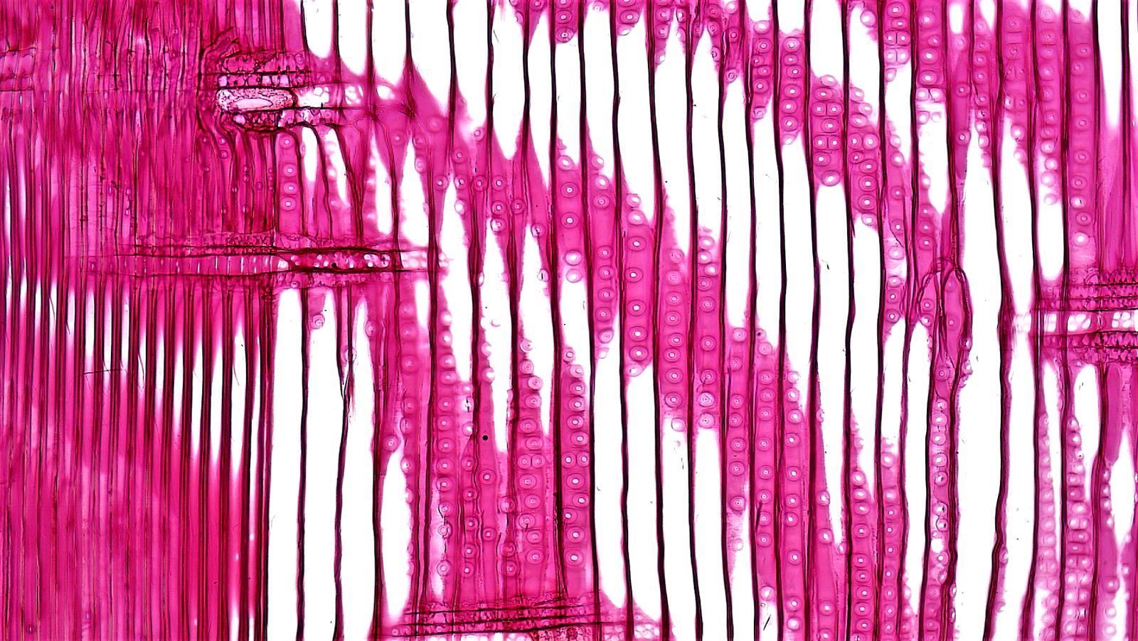
Hooke’s 50x compound microscope has descendants that would be unrecognisable to him. Confocal microscopes use laser scanning to build up three-dimensional images of living tissue. Electron microscopes, developed in the 1930s, resolve individual atoms. Super-resolution fluorescence microscopy, which won the 2014 Nobel Prize in Chemistry, breaks the diffraction limit of visible light and lets biologists watch molecular traffic inside a living cell in real time. A 2015 image of cow pulmonary artery cells, honoured in the Nikon Small World competition, shows nuclei glowing purple, mitochondria in yellow and structural fibres in blue — the interior of a single cell rendered in stained-glass colour.
The chemistry that makes those stains possible has its own long tail. The first synthetic dyes that would eventually let biologists mark specific structures inside cells came from an accidental discovery in a home laboratory over Easter 1856, when the teenage William Perkin isolated mauveine while trying to synthesise quinine. The path from Perkin’s purple sludge to fluorescent antibody stains runs through the entire history of the modern dye industry.
Even preserved biology, examined with the right instrument, gives up more than it used to. A 125-million-year-old dinosaur unearthed in China was preserved so precisely that individual skin cells remain visible under the microscope. Hooke would have understood exactly what he was looking at, and exactly what he was missing.
The word survives
Every biology textbook printed since the middle of the 19th century has repeated Hooke’s monastic metaphor, usually without noticing it. When a student in a school lab peels the skin off an onion, drops it on a slide, and turns the objective lens down to look, she is repeating the observation of 1665 with better glass. The compartments she sees are still called cells because a young man with a candle and a homemade lens thought they looked like the rooms where monks slept.
Hooke died in 1703, quarrelsome and broke. No verified portrait of him survives — some historians have suggested Newton, who outlived him and ran the Royal Society after him, saw to that. What survives is a folio the size of a small tabletop, its plates still crisp, and a single word, four letters long, doing more work in the life sciences than almost any other.