Imperial College London’s press release put the drop in clearance of a dye at “about 30% in sleeping mice”. Nicholas Franks, the Imperial biophysicist who co-led the study, said in an email that the effect was “a relatively small reduction in brain clearance during sleep (about 20%)”. The paper itself gives a third number.
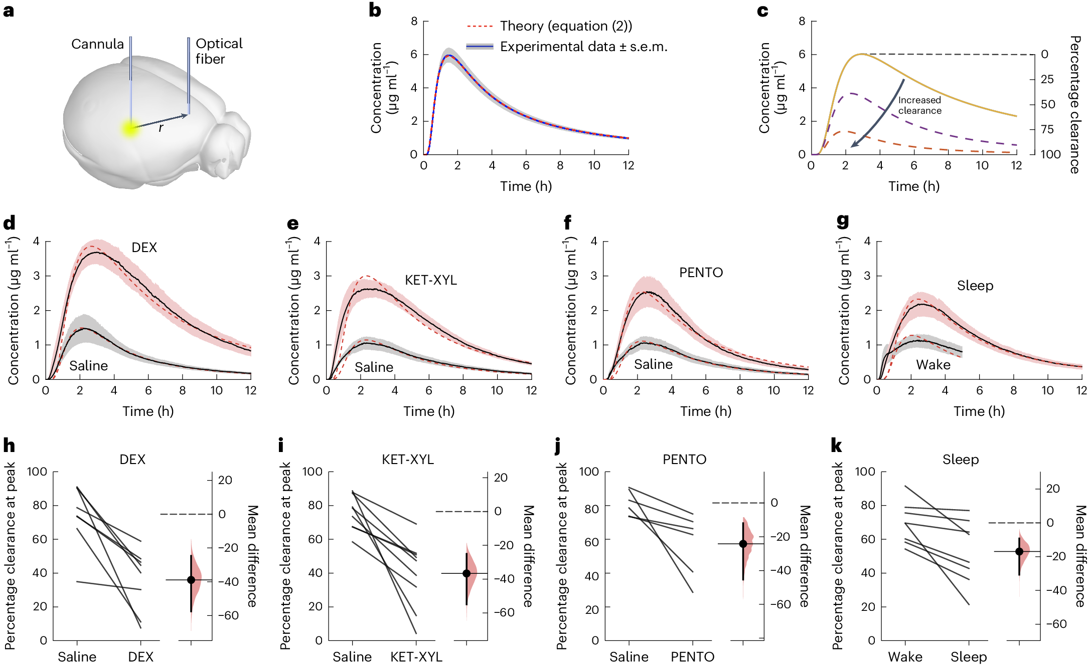
The paper, published in Nature Neuroscience in May 2024, gives the raw figures in its Extended Data Table 1. At the dye’s peak, mice kept awake had cleared 69.7% of it and sleeping mice 52.8%. Eight mice were tested, each both awake and asleep.
That is a drop of 16.9 percentage points, or about a quarter measured against the awake figure. The 30% does not seem to appear anywhere in the paper. Franks’s 20% and the table’s quarter are close, though it is not clear how he arrived at his figure.
All three numbers agree on the direction, which is that less dye was cleared during sleep in these mice. That runs against the idea that sleep washes waste out of the brain, including the amyloid-beta that builds up in Alzheimer’s disease. Underneath the percentages sits a dispute between two labs over what counts as clearance.
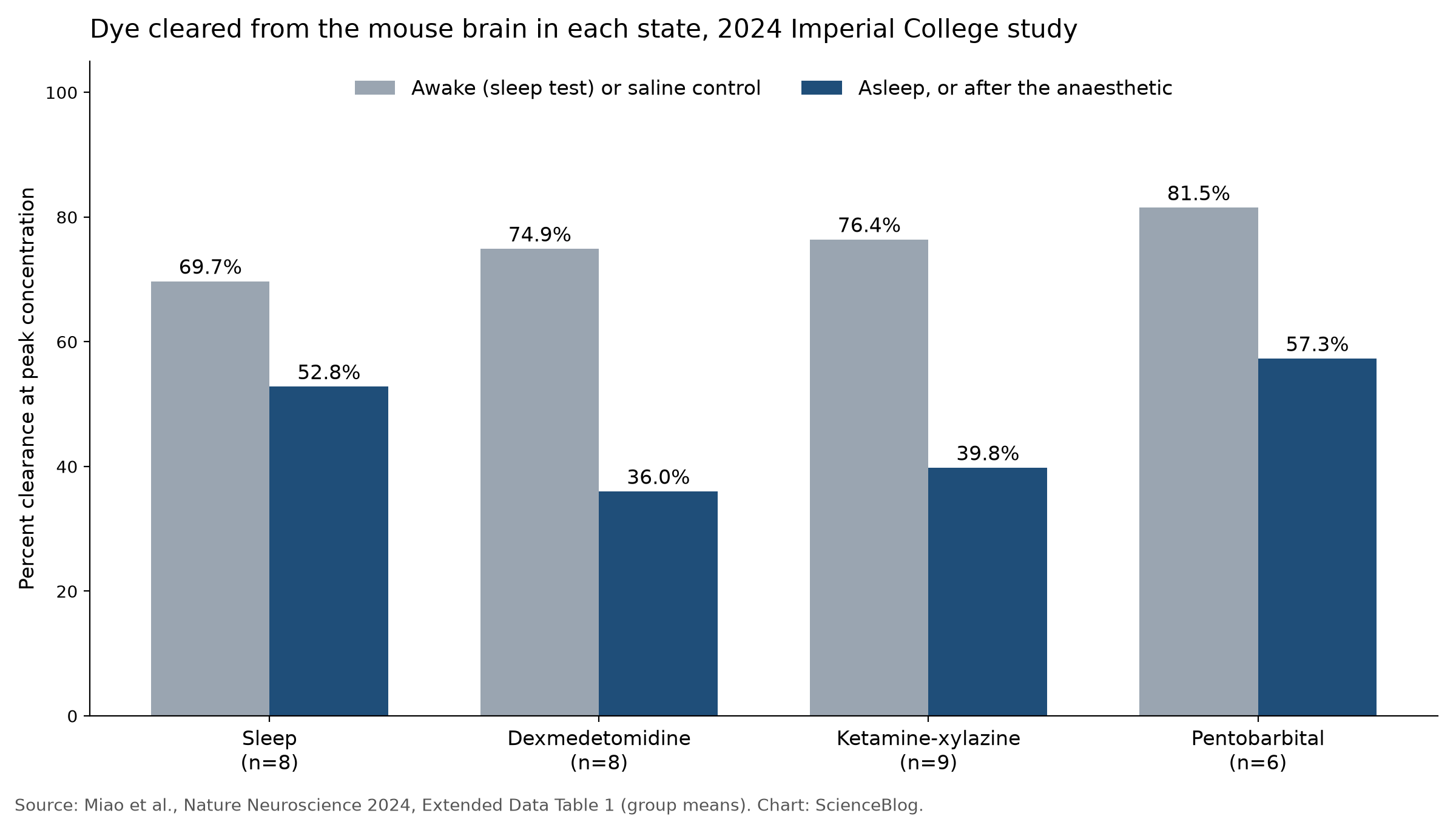
The idea Franks disputes comes from Maiken Nedergaard’s lab at the University of Rochester, which proposed that cerebrospinal fluid, or CSF, recirculates through brain tissue along paths that follow blood vessels, exchanging with the fluid between cells. The brain has no conventional lymphatic system, and in 2012 the group named this the glymphatic system.
In 2013, Lulu Xie and colleagues in that lab reported in Science that a tracer injected into the CSF of awake mice got into the brain about 95% less than in sleeping mice over half an hour of imaging. The space between brain cells grew by 60% in sleep.
When the team injected amyloid-beta straight into the cortex, the sleeping mice cleared it twice as fast. Rochester’s own press release went further than the paper, saying the glymphatic system was “almost 10-fold more active during sleep”, a figure that, as far as can be told from the paper, is not a clearance measurement.
Franks’s objection is to how the earlier work got from tracer going in to waste coming out. It “was largely based on an assumption that increased tracer entry into the brain could be used as a surrogate measurement of brain clearance,” he said in an email. “I consider this a very dubious assumption.”
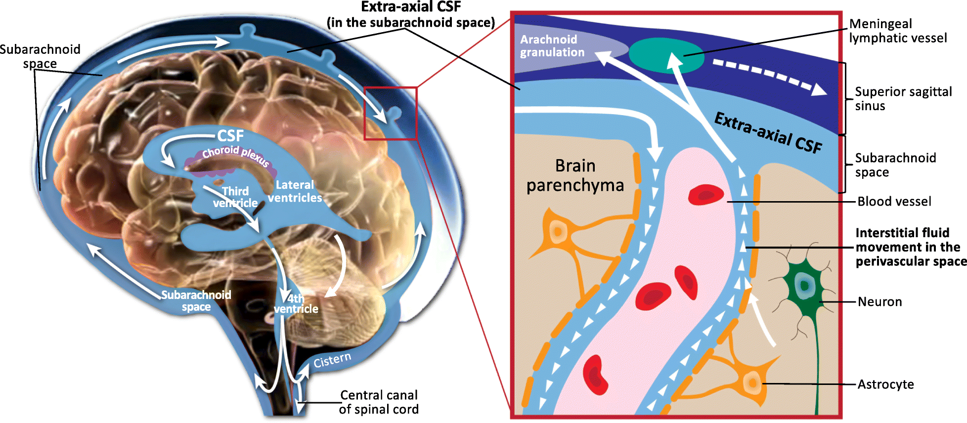
His team measured something else. Working in male mice, they injected a small fluorescent dye into a deep part of the brain, the caudate-putamen, and used an optical fibre in the frontal cortex to track how much of it arrived over the following hours, with dye lost along the way counted as cleared.
More dye arrived in the sleeping mice, which meant less had been removed, and under three anaesthetics the drop was bigger still. The authors concluded in the paper that their results “challenge the idea that the core function of sleep is to clear toxins from the brain.”
Nedergaard’s group says that design cannot show clearance at all. In a Matters Arising published in Nature Neuroscience in March 2025, Virginia Plá, Erik Kroesbergen, Nedergaard and colleagues said that dye moving from one part of the brain to another is redistribution, and that “Waste must be flushed out of the brain to be cleared.”
They also said the sleep recordings captured rebound sleep after the mice had been kept awake, that the anaesthetised mice spent most of the recordings recovering from a single dose, that the dye crosses the blood-brain barrier, and that one of the injections was too large for the tissue receiving it.
A reply from Franks and his Imperial colleague William Wisden, published the same day, called those objections “either misunderstandings, misleading, or simply incorrect”. The clearance experiments used 0.5 microlitres injected slowly, they said, and the larger volume came from separate diffusion experiments.
The 2024 authors say their result is for “a small dye that can freely move in extracellular space,” and that larger molecules may behave differently. The 2013 amyloid result came from a different experiment, with amyloid-beta itself injected into the cortex, so the two papers need not be read as one measurement giving two answers.
In January 2025, Natalie Hauglund and colleagues in Nedergaard’s lab reported in Cell that during non-REM sleep in mice, slow waves of the brain chemical norepinephrine make blood vessels tighten and relax in rhythm, pumping CSF into the brain. The sleep drug zolpidem dampened both the waves and glymphatic clearance.
The evidence in people is much thinner. In a 2021 Oslo study led by Per Kristian Eide, in which seven people stayed awake and 17 slept, one night without sleep impaired clearance of an MRI tracer from most brain regions, and later sleep did not make up for it.
A 2026 trial of 39 participants, with Jeffrey Iliff among the authors, reported higher morning blood levels of Alzheimer’s markers after a normal night’s sleep than after a night awake. The team attributed the difference to glymphatic clearance.
In mice, the two labs still disagree about how to measure clearance at all. Both human studies were small and in patients being assessed for suspected problems with CSF, and several of the 2026 authors, Iliff included, declare financial interests.