

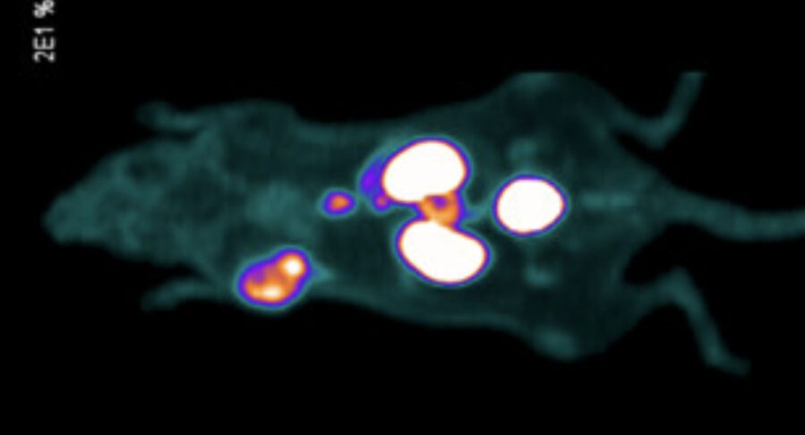
A new experimental tracer lights up prostate cancer in more ways than one. Scientists at the University of British Columbia have developed a dual-mode agent that combines PET imaging with fluorescent guidance, enabling surgeons to both see tumors and hear radioactive signals through Geiger probes.
Published in the Journal of Medicinal Chemistry, the preclinical study shows the tracer can deliver highly specific and bright tumor signals in mice with human prostate cancer implants, potentially transforming how surgeons remove cancer while sparing healthy tissue.
Prostate Cancer Surgery Needs Better Tools
Prostate cancer is among the most common cancers in men, affecting one in eight Canadians in their lifetime according to the Canadian Cancer Society. Surgery to remove tumors often involves balancing complete removal against preservation of nerves and organs like the bladder or bowel. Current tools such as PET scans and blue dyes offer guidance but are limited in precision and consistency during operations.
A Dual Mode Approach
The UBC team designed a tracer that targets prostate-specific membrane antigen (PSMA), a protein abundant on prostate cancer cells. The agent incorporates:
- Fluorine-18 labeling for high-resolution PET imaging.
- Fluorescein dyes for bright optical visualization without special equipment.
- PSMA-targeting scaffolds to ensure tumor specificity.
This dual design means surgeons could locate hidden metastases or lymph nodes by sound with a handheld radioactive probe, while also seeing fluorescence in the tumor tissue itself.
“Our tracer provides high-resolution visual guidance, but would also allow a surgeon to use a hand-held Geiger counter probe to ‘hear’ areas of high radiation density that would accumulate in cancerous tissue not immediately visible,” said Dr. David M Perrin, a UBC chemist and senior author (Journal of Medicinal Chemistry).
Preclinical Results And Expert Reactions
The study, conducted with colleagues at BC Cancer, tested the tracer in mice with implanted human prostate tumors. Results showed strong uptake in tumors (7–17% injected dose per gram) and bright fluorescence both in cell cultures and tissue samples. First author Jerome Lozada noted that the compound’s simple chemistry makes it highly translatable even for smaller hospitals that rely on standard PET and surgical equipment.
Outside experts highlighted the potential impact. Dr. Larry Goldenberg of the Vancouver Prostate Centre said dual mode tracers could reduce surgical complications by helping surgeons avoid damaging critical structures while still ensuring thorough tumor removal. Nuclear medicine specialist Dr. Philip F Cohen of Lions Gate Hospital compared the approach to established breast cancer tracers, but emphasized the benefit of combining functions into a single injection.
Next Steps Toward The Clinic
The team is now preparing the tracer for Good Manufacturing Practices review, toxicity studies, and validation trials. If successful, the approach could be applied to other cancers where both radioguided and fluorescence-guided surgery offer advantages, including ovarian and laryngeal cancers.
As Perrin’s team works toward clinical testing, the innovation reflects a growing trend in precision oncology: combining diagnostic imaging and surgical navigation into streamlined, patient-friendly tools. With more consistent detection of tumor margins and metastatic spread, dual mode tracers could improve survival while reducing side effects.
Journal: Journal of Medicinal Chemistry
DOI: 10.1021/acs.jmedchem.5c01480