

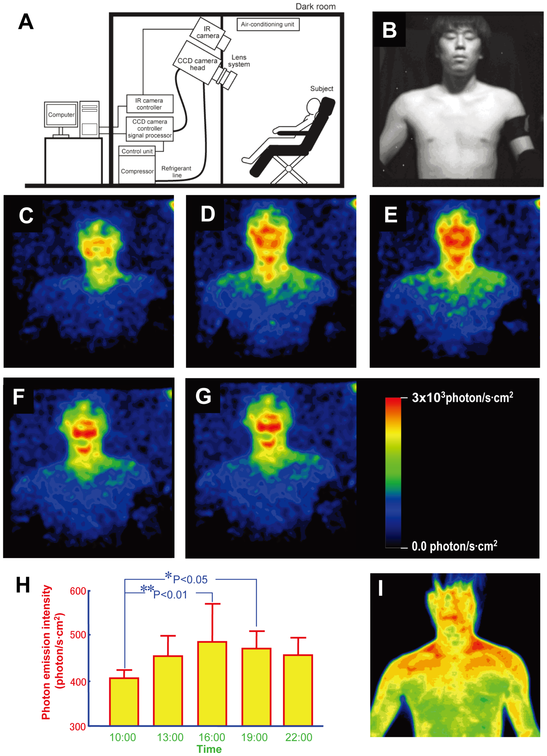
A room at the Tohoku Institute of Technology in Sendai was sealed against outside light. One by one, five men entered it, sat bare-chested in front of a camera and waited 15 minutes in complete darkness. Then they held still for a 20-minute exposure.
The pictures that emerged look like scenes from science fiction. Blue, green and red contours cover the body, with the strongest concentrations around the face. But nobody in the room could see that light. The colours were added to represent intensity, and the signal itself was about 1,000 times fainter than the threshold the researchers gave for unaided human vision.
What the camera measured was nevertheless real light. The photons fell within wavelengths that human eyes can ordinarily detect when enough of them arrive. The experiment’s achievement was to gather an extremely sparse stream of those photons into an image and then follow how it changed across the day.
The resulting 2009 PLOS ONE paper, led by Masaki Kobayashi, offers a useful lesson in how an accurate fact can become misleading when its scale disappears. Human bodies emit visible light. That does not mean humans are visibly luminous.
Twenty minutes in darkness
The five participants were healthy men in their twenties with no skin conditions and no oral medication. The researchers asked them to keep regular sleep schedules for a week before the experiment, generally sleeping from midnight until 7 am under normal light and dark conditions.
On a measurement day, each man waited in a neighbouring room illuminated to about 400 lux. He removed clothing from the upper body, did not use cosmetics or aftershave, and lightly wiped his skin with lukewarm water before entering the dark chamber alone.
There he sat in front of the detector, listening to comfortable music without falling asleep. The team allowed 15 minutes of dark adaptation, then opened the camera’s shutter for 20 minutes. They repeated the process every three hours from 10 am to 10 pm, across three days.
At each session the researchers also collected saliva for cortisol analysis, measured oral temperature and took a thermal image. Those parallel measurements helped them ask whether the changing photon count simply mirrored heat or followed some other daily biological rhythm.
This was a tightly controlled demonstration, not a survey of humanity. Five young men are too few to show how emission might differ in women, older people, children, illness, different skin types or different seasons. The narrow sample is important whenever the striking images are presented as a universal portrait of the human body.
The camera was cold because the signal was faint
The detector was a cryogenic charge-coupled device, or CCD, cooled to minus 120 degrees Celsius. At ordinary temperatures, electrical activity within a sensor creates noise. Cooling it so severely reduced that background enough for the instrument to distinguish an exceedingly weak biological signal.
The camera had the ability to detect light at the level of a single photon. That description is accurate, but it can leave the wrong impression. The published image was not a snapshot made from one miraculous photon. Each frame integrated detections over a full 20 minutes, allowing a spatial pattern to emerge from light that arrived too sparsely for an eye to recognize.
The raw detector produced a numerical map. Researchers then assigned colours to different intensities. Red indicated a relatively high photon count and blue a lower one. Those colours did not show the hue of the emitted light, and the outline was not what a dark-adapted observer would have seen.
Earlier attempts to image ultraweak human emission could require exposures longer than an hour. The improved sensitivity mattered because a camera that needed most of an afternoon to form one image could not resolve meaningful changes at several points in the same day.
Visible wavelengths do not mean visible bodies
Previous spectral measurements discussed in the paper placed human ultraweak photon emission broadly between 500 and 700 nanometres, with reported peaks in green-yellow and red portions of the spectrum. Those wavelengths fall within the band called visible light.
Visibility, however, depends on more than wavelength. A sound can fall within the frequencies humans hear and still be too quiet to notice. Light works similarly. A photon may carry a visible wavelength while the total stream remains far below conscious perception.
Under carefully optimized laboratory conditions, dark-adapted volunteers have sometimes reported a single photon aimed at a sensitive part of the retina. That does not mean every isolated photon becomes a visible flash. Perception is probabilistic and depends on timing, location, optical losses and the brain’s decision threshold.
The body presents a different problem. Its photons are scattered over a large area and emitted slowly, rather than delivered as a controlled pulse to one retinal location. The 1,000-fold figure in the 2009 paper is therefore a useful order-of-magnitude comparison, not the result of asking the five men whether they could see one another glow.
This is also why “biophoton,” a term sometimes attached to unsupported claims about auras or healing energy, needs care. In this experiment it meant measurable ultraweak photon emission from biological chemistry. The study did not photograph a mystical field, test whether mood changes its colour or demonstrate that the light carries messages between people.
The face brightened in late afternoon
The most interesting result was not simply that photons existed. It was that their number changed with time. Emission from the face and upper body was relatively weak at 10 am, rose through the afternoon, reached its highest measured level around 4 pm, then declined toward 10 pm.
The face generally produced more photons than the torso. The mouth and cheeks were especially prominent. In a cheek region selected for analysis, output at the afternoon maximum reached roughly 3,000 photons per second per square centimetre, about twice the level measured in the morning.
Salivary cortisol followed an opposing curve and had a statistically significant negative correlation with photon emission. The researchers interpreted this as evidence that the light could reflect a circadian rhythm in metabolic activity. Oral temperature did not show the same relationship.
Three additional sleep-deprived volunteers were measured at 1, 4 and 7 am under constant light conditions, and their emission remained low overnight. That extra test supported the daily-rhythm interpretation, but three people are even less sufficient for broad conclusions. It did not reveal which clock-controlled chemical reactions created the curve.
The timing also means the body’s brightest measured period should not be confused with the warmest-looking region in a thermal picture. When the researchers placed the photon map beside infrared thermography, the two images looked different.
This was chemistry, not body heat
Every object above absolute zero emits thermal radiation. A human body produces abundant infrared light because of its temperature, which is why a thermal camera can show a person in darkness. That familiar effect was not what the cryogenic CCD was measuring.
The study found no significant correlation between oral temperature and photon intensity. Its thermographic distribution also failed to match the visible-wavelength photon map. The apparent red areas in the paper’s emission images were high values on a false-colour scale, not infrared warmth and not red light seen by the participants.
The leading physical explanation begins with oxidative metabolism. Normal cellular chemistry generates reactive oxygen species and excited molecules. When some of those molecules return to a lower-energy state, a tiny share of their energy can leave as photons.
This is spontaneous ultraweak emission, not the specialized enzyme-driven bioluminescence that lets a firefly flash. It is closer to a faint by-product of chemistry than a biological lamp. The face may be brighter partly because of differences in skin metabolism, blood flow, pigment and exposure to light, but the experiment did not isolate one cause.
Nor did it establish that the photons perform a biological function. Measuring light from a reaction is not evidence that cells use that light to communicate. A detector can reveal exhaust from a process without showing that the exhaust is a signal.
What the picture proves, and what it leaves open
The Sendai experiment was not the first evidence that living tissue emits ultraweak photons. Its important advance was speed. By reducing exposures to 20 minutes, the researchers could image people often enough to reveal a possible daily rhythm rather than average that rhythm away in a single hour-long observation.
Research has continued. A 2025 study of mice and plants reported stronger ultraweak emission in living mice than after death, despite keeping temperature controlled, and changes in plant leaves exposed to heat or injury. The results extended the case that oxidative state and biological stress can alter the signal, while leaving its possible uses and mechanisms under investigation.
As ScienceBlog noted in its coverage of that later work, highly sensitive cameras can make this emission legible without making it visible to us. The instrument is not exposing a hidden view that human eyes would see if only the room were darker. It is accumulating and translating a signal across time.
A larger modern study could test women and men across ages, repeat measurements in different seasons, record full spectra and control sleep, meals, exercise and light exposure. It could also examine whether the 4 pm peak holds across different circadian schedules or merely described this small group under one protocol.
Until then, the 2009 images deserve both wonder and restraint. Ordinary human chemistry releases occasional visible-wavelength photons. A chilled sensor, a sealed room and 20 minutes of patience can count them. None of that grants us luminous outlines in the dark.
The light is real. The luminous person our eyes imagine is not.