
Inside a developing child, the brain never experiences time alone. Its cells are exposed to blood flow, hormones, nutrients, immune signals, movement and a growing stream of sensory activity. Every moment is embedded in the rest of the body.
That is what makes a long-running laboratory experiment so revealing. Peppercorn-sized clusters of human cortical cells, grown without a body and without eyes, ears or other sensory organs, continued to pass through part of the molecular sequence seen during human brain development.
In a study published in Nature on 19 August 2026, a Harvard- and Broad Institute-led team tracked these cerebral cortex organoids across an interval that once seemed impractical. The peer-reviewed molecular time series reached five years. Neurons were detected in intact tissue at 5.8 years, and a Harvard and Broad account says the laboratory had some cultures that were seven years old.
Those are three related but different claims. Seven years describes the survival of some cultures, not seven years of identical molecular sampling. The paper’s most comprehensive measurements stop at five years. What they reveal is not merely tissue that remained alive, but tissue that kept changing on a recognisably human schedule.
A peppercorn is not a miniature brain
The organoids began with human pluripotent stem cells. Some lines were produced by reprogramming mature donor cells, while the study also used a human embryonic stem-cell line. The researchers placed thousands of cells in low-attachment wells and applied biochemical signals that steered them toward a dorsal cerebral cortex identity.
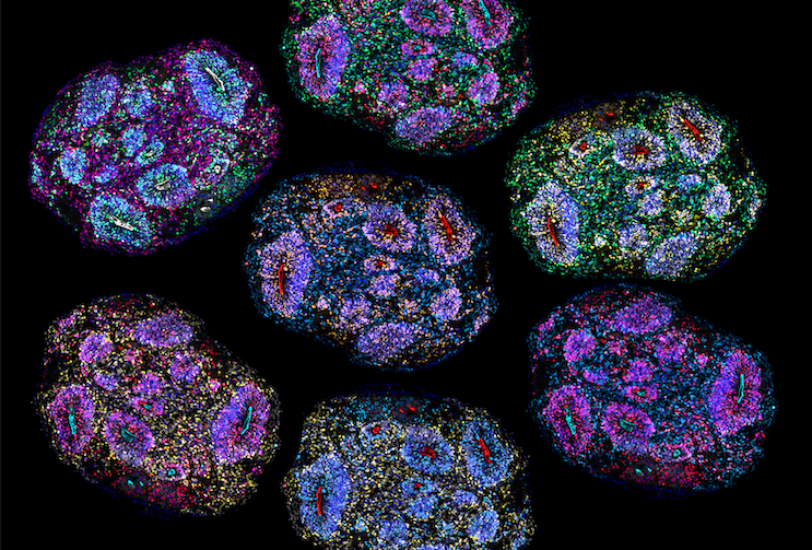
The resulting spheres contained more than one million cells apiece, according to the Broad Institute. They produced neural progenitors, several kinds of neurons, astrocytes and other cell populations associated with developing cortex. Their three-dimensional form let cells organise and connect in ways that flat layers in a dish cannot easily reproduce.
But “brain organoid” names a model, not a small complete organ. These clusters did not reproduce the full anatomy of a brain, attach to a body, receive natural sensory input or participate in behaviour. They had no skull, circulation or lived experience. Nothing in the study establishes perception, consciousness or thought.
This boundary has long mattered in organoid reporting. ScienceBlog has also examined organoids connected to electronic systems for game-like tasks. Those preparations receive deliberately encoded inputs and feedback. The long-lived organoids here were being used to model development, not to play a game. Scientists construct tractable pieces of biology so that processes normally hidden inside pregnancy and childhood can be observed directly.
The time series followed 424,720 individual cells
Irene Faravelli, Noelia Antón-Bolaños and their colleagues newly analysed 34 individual organoids collected at six months, nine months, and one, 1.5, two, three, four and five years. They integrated these samples with 76 organoids from earlier work, beginning at 15 days and continuing through six months.
The combined atlas contained 110 organoids and 424,720 cells across 16 timepoints. For the stages from six months to five years, the series included at least six organoids drawn from three experimental batches. That replication matters because organoids can vary from one preparation to another, and different cell types do not survive long culture equally well.
Single-cell RNA sequencing showed which genes were active in each recovered cell. The researchers then compared those patterns with reference datasets from human cortex. Early organoids, from 15 days to two months, mapped most closely to first-trimester tissue. Three- to six-month cultures generally resembled the second trimester. From nine months onward, the molecular profiles shifted progressively toward late-prenatal and postnatal ages.
Postnatal-like signatures began emerging after about 12 months in culture. This does not mean a one-year-old organoid was equivalent to a one-year-old child’s brain. It means that particular cell populations and gene programs had moved into ranges associated with development after birth.
DNA carried a second record of age
Gene activity is only one way to estimate biological time. The team also measured DNA methylation, chemical marks that accumulate or disappear at particular locations in the genome. Some age-associated sites can be combined into “epigenetic clocks” trained on tissue of known age.
Between three months and five years, whole-genome analysis identified 213 regions whose methylation changed with organoid age. Many occurred near regulatory features and genes important to development of the neocortex. The organoids also accumulated a form of non-CpG methylation considered a marker of neuronal maturity.
When the researchers applied established clocks, the predicted DNA-methylation age tracked time in culture closely. Correlations were 0.88 for one model and 0.90 for another. Their median absolute errors were 7.25 months and 20.04 months, respectively. A fetal-brain clock produced a weaker correlation of 0.54 and narrowly missed conventional statistical significance. The NIH summary of the work describes this agreement as a lifelike developmental pace.
These details prevent “molecular clock” from becoming mystical. The cells did not count days. Their genomes moved through coordinated chemical states that human reference data allow scientists to read as age. The match was strong in two models, imperfect in all of them and dependent on which clock was used.
Survival did not mean every cell aged equally well
The team detected the expected broad classes of cortical cells across the series, but their proportions changed. Neurons are especially fragile during long culture and can also be lost when tissue is dissociated for single-cell sequencing. By later timepoints, glial cells were more prominent in the recovered molecular data.
To check intact tissue directly, the researchers stained organoid slices rather than separating them into cells. A general neuronal marker remained visible at two, three, four and 5.8 years. SATB2, a marker used here for a class of excitatory cortical projection neurons, was also present at 5.8 years.
A specially designed activity-permissive medium helped excitatory neurons survive and mature. In that medium, the cells developed more elaborate shapes and synaptic structures. The organoids generated spontaneous electrical activity that researchers followed for at least two years.
Electrical signals do not turn the tissue into a thinking brain. Neurons are electrically active cells, and coordinated activity is one property a useful neural model should reproduce. Separate ScienceBlog coverage of structured electrical sequences forming without sensory experience illustrates how activity can self-organise in immature neural tissue. It still does not establish thought or awareness.
Older progenitors remembered which steps had passed
The strongest evidence for an internal timetable came from an experiment that disturbed the normal sequence. The team took progenitor cells from organoids aged nine to twelve months, separated them and reassembled them either alone or alongside progenitors from 15-day-old organoids.
After two weeks, the young cells produced the early cell types expected near the beginning of cortical development. Older progenitors sharing the same new environment produced later-born cortical projection neurons, glial precursors and astrocytes. Some of those fates normally take two or three months to appear in culture.
The old cells were not rigid. Signals from their young neighbours pushed them back toward neurogenesis. Yet they did not restart the entire sequence. They responded from the developmental position they had already reached, skipping early products and generating later ones.
The researchers call this the ability to record and recall developmental time. “Memory” here is cellular, not autobiographical. It refers to a durable molecular state that changes which descendants a progenitor makes. No experiences were stored and no organoid remembered an event.
The clock is intrinsic, but it is not independent
Human neurons mature unusually slowly even when removed from a person. The new results add evidence that some of this pace is cell-intrinsic: biological machinery within human cells constrains the order and speed of developmental change.
Intrinsic does not mean sealed off from the environment. The activity-permissive medium changed neuronal survival and maturation. Young cells altered the behaviour of old progenitors. Oxygen stress remained concentrated near the organoids’ cores, where the absence of blood vessels makes diffusion difficult. The cultures carried a timetable, but conditions influenced which parts of it could unfold.
The paper itself points to what is missing. Natural brain development is shaped by evoked activity from sensory systems and by non-neural tissues throughout the body. Future organoids may incorporate more of those influences, but adding complexity will not make every feature of an organoid equivalent to a feature of a person.
The distinction is scientifically useful. If development were entirely imposed from outside, isolated cells should quickly lose their schedule. If it were entirely autonomous, changing their medium or neighbours should not matter. The experiment instead reveals an internal sequence continually negotiated with local conditions.
Five years opens development that was previously inaccessible
The human cerebral cortex takes close to two decades to mature. Mice and other laboratory animals develop on much faster schedules, while direct sampling of healthy human brain development is severely limited. Most organoid experiments have therefore captured prenatal or very early stages over a few months.
The previous published longevity benchmark followed cortical organoids for as long as 694 days. That work showed key early postnatal transitions. Extending reproducible cultures beyond five years gives researchers access to later cell states and a way to ask when genetic differences linked to autism, epilepsy, schizophrenia or neurodegeneration first alter development.
It does not guarantee that a disorder can be recreated accurately or that a treatment found in a dish will work in a person. Long cultures are expensive, slow and technically fragile. The Harvard team is now interested in using the temporal-memory result to reach selected later stages faster, rather than maintaining tissue simply to break another longevity record.
The experiment’s quiet surprise is not that scientists grew a brain in a dish. They did not. It is that human cortical cells, separated from nearly everything that usually marks the passage from gestation into childhood, still carried enough of their developmental history to know approximately when they were.