
Silence was supposed to be the blank condition. Instead, it produced the one result that lasted.
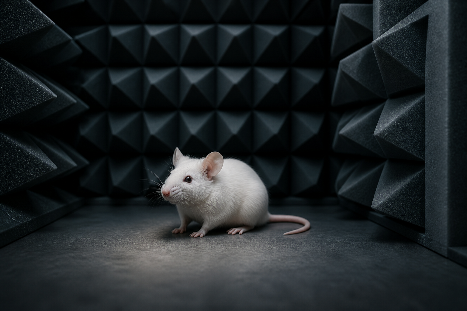
In a 2013 study, young female mice spent two hours a day inside an anechoic chamber. Some heard white noise, recorded pup calls or Mozart’s Sonata for Two Pianos in D major. One group heard only the residual quiet the chamber could provide. When the researchers examined the hippocampus seven days after labeling newly dividing cells, only the silence group had more surviving labeled cells than mice kept with ordinary animal-facility sound.
The experiment was small, conducted in mice and focused on cell markers. It included no test of memory, learning, mood or behavior. This is one mouse study, not settled consensus about what silence does to brains, and it provides no evidence that two quiet hours will cause the same cellular change in a person.
“Silence” meant an unusual laboratory environment
Imke Kirste, Zeina Nicola, Golo Kronenberg, Tara Walker, Robert Liu and Gerd Kempermann reported the work in Brain Structure and Function. The paper appeared online on December 1, 2013, although it was assigned to the journal’s March 2015 print issue.
The subjects were female C57BL/6J mice, six to eight weeks old at the start. Experimental groups contained ten animals. The researchers chose this strain partly because it has relatively high neurogenic activity in the dentate gyrus, the hippocampal region they planned to count. Because this strain can develop hearing loss later in life, they separately measured auditory brainstem responses in young mice and confirmed that the planned sounds were audible at the study age.
Each exposure began near the start of the animals’ dark phase and lasted two hours. The mice entered an anechoic sound-isolation box designed to absorb reflections and keep outside noise from interfering. The “silence” condition was therefore not a claim of physically perfect, zero-decibel silence. It meant isolation from the experimental sounds and most of the facility’s normal auditory background.
The comparison condition was ordinary animal-house noise. Four other groups experienced the anechoic chamber with white noise, mouse pup calls, Mozart or silence. White noise covered roughly 4 to 80 kilohertz at 70 decibels. The pup calls averaged around 65 kilohertz. The Mozart recording was filtered and shifted five octaves upward so most of its power fell between 5 and 20 kilohertz, within a useful range for the mice.
That frequency shift matters. The experiment did not ask whether mice appreciated human classical music as humans hear it. Mozart supplied a complex, patterned sound without known biological relevance to the animals.
The paper ran two timelines, not one
The researchers wanted to distinguish an immediate increase in precursor-cell division from the early survival and differentiation of the resulting cells. Those are different stages, so they used two BrdU schedules.
Bromodeoxyuridine, usually shortened to BrdU, is a synthetic building block that cells incorporate into newly made DNA. An injection creates a time stamp for cells passing through DNA synthesis around that period. Later staining can reveal where those labeled cells are and which other molecular markers they carry.
For the proliferation protocol, mice received two-hour exposures on three consecutive days. The BrdU injection came 24 hours after the last exposure, and the brains were collected another 24 hours later. This short interval asked whether the recent auditory environments had changed the number of dividing precursor cells.
White noise did not produce a detectable difference from ambient facility sound. Pup calls, Mozart and silence all did. Each was associated with more BrdU-positive cells in the dentate gyrus, with the highest counts in the silence and Mozart groups. Those two conditions also increased BrdU-labeled cells carrying Sox2, a protein used here to identify early neural precursor stages.
So Mozart did have an effect in this study, but it was a short-term proliferation result. The title’s “silence, not Mozart” distinction belongs to the later measurement.
At seven days, silence was the only condition still different
For the survival-and-differentiation protocol, the order changed. Mice received BrdU first, then underwent two hours of their assigned auditory condition on seven consecutive days. Researchers collected the brains 24 hours after the final exposure. The labeled cells were therefore about seven days into their development.
By that point, the groups exposed to Mozart, pup calls and white noise were statistically indistinguishable from the ambient-noise controls in total BrdU-positive cell counts. The silence group alone retained an increase. Across all five conditions, the overall difference was strong in the authors’ analysis.
The team then examined the identity of labeled cells in the silence and ambient groups. NeuN staining was used as a neuronal marker, while S100β identified astrocytes. Roughly two-thirds of labeled cells were neurons and around 15 percent were astrocytes.
The neuronal share was 62 percent in the silence condition and 57 percent with ambient noise. That difference in proportions narrowly missed the conventional statistical threshold, with a reported p value of 0.056. In absolute numbers, however, silence produced significantly more cells carrying both BrdU and NeuN, with p equal to 0.008.
This is the precise basis for saying the silence group had more seven-day-old immature neurons. BrdU marked cells born around the labeling window, and NeuN supported their neuronal identity. The experiment did not watch mature neurons appear from nothing during two quiet hours.
Seven-day survival is not the same as a working memory circuit
At seven days, adult-born hippocampal cells are still early in a long developmental process. They must continue surviving, extend processes, receive and send synaptic input, and integrate into an existing circuit. Many newly generated cells do not complete that path.
The researchers described their later BrdU count as an early-survival measure. They did not follow the cells for weeks or months, test their electrical integration, or establish that the numerical difference persisted. “Increased neurogenesis” in the paper refers to more BrdU/NeuN-positive cells at this early stage, not a demonstrated permanent expansion of the functioning hippocampus.
The same boundary applies to cognition. The hippocampus contributes to memory and spatial processing, and adult-born dentate-gyrus neurons have been investigated for roles in distinguishing similar experiences. But a plausible role for a cell population is not evidence that changing its early count improved a mouse’s performance.
No animals in this experiment ran a memory maze, completed a recognition task or received any other behavioral assessment. The study cannot tell whether the silence group learned faster, remembered longer or behaved differently at all.
It also cannot settle whether the same response exists in humans. The experiment used one young inbred mouse strain under a highly controlled auditory protocol. Human environments, developmental stages and hippocampal cell dynamics are not interchangeable with that model.
Why an absence of sound might behave like a stimulus
The result looks paradoxical only if silence is treated as nothing. For mice accustomed to the constant background of an animal facility, an anechoic chamber may be an unusually salient environment. Removing expected sensory input can itself create novelty.
Kirste and colleagues proposed that novelty could help explain why silence and the frequency-shifted Mozart initially produced large precursor responses. Both were unfamiliar, while pup calls were biologically familiar and white noise was unstructured. On their account, the absence of sound might increase alertness as the animal monitored a changed environment.
The authors also considered the possibility that silence permits internally generated brain activity rather than simply reducing stimulation. But these were interpretations, not mechanisms demonstrated by the experiment. The study did not directly measure alertness, stress hormones, auditory-cortex dynamics or the subjective state of the mice.
Nor did it separate quiet from every feature of the chamber experience. The control groups also entered the anechoic box, which helps, but a silent laboratory interval still differs from a naturally quiet nest in temperature, social cues, handling and expectation. “Silence” is the name of an experimental condition, not a universal biological dose.
A cell-count result, not a prescription
The careful reading is more interesting than the viral version. Several structured auditory conditions briefly increased precursor proliferation. Only silence was associated with a larger surviving labeled population seven days later, including more cells carrying a neuronal marker. That difference invites a question about how unexpected sensory absence affects early hippocampal plasticity.
It does not show that Mozart is ineffective for every brain outcome, that white noise damages neurons, or that silence makes an animal smarter. The experiment was not designed to compare wellbeing, attention, sleep or learning. Its five conditions answer one narrow cellular question under one protocol.
There is therefore no evidence here for prescribing two silent hours a day. The duration was an experimental exposure chosen for mice, not a tested human routine. A person seeking quiet may value it for ordinary reasons without attaching an unmeasured neurogenesis claim.
What survived the experiment was a modest but clear surprise: the condition intended to remove auditory stimulation became the only condition associated with more seven-day-old immature hippocampal neurons. The study counted cells, not memories, and that is exactly where its conclusion should stop.