

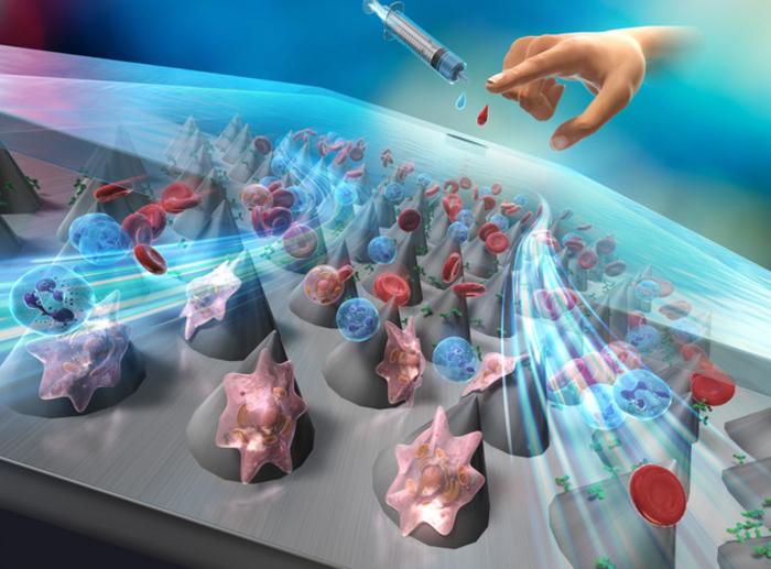
A team of engineers from Chiba University in Japan has created a simple yet highly sensitive tool that captures cancer cells from blood samples, using a device made from embossed plastic and coated antibodies.
Their microfluidic system, described in the journal Lab on a Chip, traps circulating tumor cells (CTCs) with more than 90% efficiency, even at high flow rates. This approach could pave the way for affordable, minimally invasive blood tests to detect cancer and monitor recurrence after treatment.
How the Device Works
Circulating tumor cells are rare and notoriously difficult to isolate from blood. These cells shed from primary tumors and may seed metastasis elsewhere in the body. While microfluidic technologies have been used to trap them before, they often require expensive, complex manufacturing steps. The new device overcomes these hurdles by using polycarbonate (PC) sheets embedded with cell-sized microcones, produced through thermal nanoimprint lithography (T-NIL).
Each microcone is about 30 micrometers wide and tall, arranged in a tightly packed hexagonal pattern. The textured surfaces of these cones strongly attract antibodies without any chemical linkers. Researchers coated them with antibodies that recognize EpCAM, a protein found on many cancer cells.
Simple Assembly, Strong Performance
Instead of building complex channels, the team formed a “microgap” channel by sandwiching the PC sheet between a glass slide and a soft polymer plate. Blood is then pumped through this gap. As cancer cells tumble through, they brush against the antibody-coated cones and stick.
- More than 90% of breast cancer cells (MCF-7) were captured at flow rates up to 100 µL/min
- The optimal cone angle was 15° or 30°, tilting cones toward the flow
- The device removed over 99% of non-cancerous white blood cells
- Cancer cells remained fixed in place during multi-step fluorescent staining
- Capture also worked for lung cancer cells (A549), though not for EpCAM-negative cervical cancer cells (HeLa)
“There are many technologies for detecting cancer, but it has been a long-standing challenge to detect cancer cells with high sensitivity using minimally invasive methods,” said Professor Masumi Yamada, the study’s lead investigator.
Why Cone Orientation Matters
To maximize performance, the researchers adjusted the angle of the microcone array relative to the flow direction. At 0°, most cells passed straight through. But when tilted at 15° or 30°, cells collided more often with the cones, boosting capture efficiency. High-speed flow imaging confirmed that tilted arrays disrupted the smooth flow and increased particle-cell interactions.
“Cells would have an equal opportunity to collide with microcones regardless of their initial position,” the team noted, citing their confocal particle tracking studies.
Built for Real-World Diagnostics
Unlike many CTC-capture devices that rely on intricate chemical modifications or soft polymers like PDMS, this system uses mass-producible polycarbonate sheets. The surface doesn’t need elaborate preparation and can hold antibody coatings for over a year. After trapping cells, researchers successfully performed in situ staining within the channel, highlighting the device’s compatibility with downstream analysis.
In future clinical use, such systems could be multiplexed to capture different cancer cell types by combining antibodies against markers like HER2 or CD44. The team also plans to test patient-derived blood samples, simulate fluid dynamics in silico, and scale up throughput.
A Path Toward Early, Accessible Cancer Screening
The simplicity of this device could transform how we monitor cancer—both in terms of early detection and checking for recurrence after treatment. A test that only requires a small blood sample, processed through a low-cost, high-efficiency plastic chip, might one day be part of routine cancer screening.
Journal: Lab on a Chip
DOI: 10.1039/d5lc00143a
Article Title: Enhancing cancer cell immunocapture on orientation-controlled nanoimprinted microcone arrays in microgap channels
Publication Date: May 28, 2025