
Water is the enemy. In the terahertz band, that slice of the spectrum wedged between microwaves and infrared light, water swallows the radiation more than a million times more greedily than it swallows visible light. Shine a terahertz beam at living tissue, which is mostly water, and most of it never comes back. For thirty years this has been the wall that terahertz imaging kept running into. And yet the same thirsty absorption that ruins the picture is, it turns out, the very thing that could let these waves read the body in ways ordinary light cannot.
That paradox sits at the heart of a new review by Kazunori Serita at Waseda University and Masayoshi Tonouchi at Okayama University, both in Japan, published in the Journal of Physics Photonics. Terahertz biophotonics, they argue, has spent two decades stuck between high hopes and stubbornly few real uses.
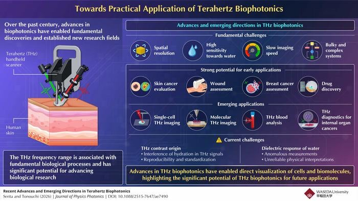
The hopes are easy to understand. Terahertz waves are non-ionising, so they will not damage DNA the way X-rays can. They pass through some tissues with less scattering than visible light because their wavelength is longer. And they are exquisitely tuned to the wobble and rotation of molecules, picking up “fingerprint” signatures that betray what a substance actually is. The snag is everything else. Resolution is coarse because the wavelengths are long. The hardware is bulky, the imaging slow, and water, as ever, gets in the way.
“THz biophotonics is a fascinating research area for next-generation biomedical technologies,” says Serita. “Currently, THz biomedical applications are restricted to a few niche domains with many technical limitations. Recent developments in emerging THz technologies have greatly increased the potential for overcoming these technical limitations.”
Reading content, not shape
Here is the clever reframing. Most medical imaging, from the dermatologist’s magnifier to the MRI scanner, looks at structure. Terahertz looks at water. Cancerous tissue tends to be more hydrated than the healthy stuff around it, swollen with the extra fluid that comes with rapid cell growth, inflammation and new blood vessels. So a tumour that hides from the eye can light up in terahertz, not because of how it looks but because of how wet it is. Serita and Tonouchi put it as probing the “content” of a lesion rather than its “shape”, and that is a genuinely different kind of contrast from anything an optical microscope offers.
Skin has been the obvious place to start, being right there on the surface where water absorption matters least. Skin cancer diagnosis still leans on an uncomfortable rule of thumb: when in doubt, cut it out. Plenty of benign moles get excised just in case. Terahertz might trim that uncertainty, and the work has crept from the lab bench toward actual patients. One group built a robot-assisted handheld probe, charmingly named the PicoBot, that controls exactly how hard it presses on the skin and at what angle, then took it into a university hospital to scan real skin cancer patients.
Wound care, oddly, may be even closer to the clinic than cancer. Because a burn or a healing scar is fundamentally a story about hydration, and terahertz reads hydration like nothing else, the technology slots in neatly. Researchers have used handheld terahertz scanners on burns in animal models, sorting partial-thickness burns by severity against the histological gold standard, and even feeding the dielectric measurements into neural networks to predict whether a wound will heal on its own or need intervention. For diabetic foot syndrome, where dried-out patches of sole skin flag where ulcers may form, terahertz hydration maps have lined up with clinical assessments of nerve damage. That is the sort of quiet, practical win that rarely makes headlines but matters enormously to the people it helps.
The breakthrough is a point of light
What changes the game, the review suggests, is a clutch of new ways to make and aim the waves. The standout is the scanning point terahertz source, or SPoTS, microscope. Instead of flooding a sample with a broad terahertz beam and fighting the diffraction limit, SPoTS focuses a femtosecond fibre laser to a tiny spot on a special crystal, generating a terahertz source smaller than the terahertz wavelength itself, right next to the sample. The upshot is micrometre-scale resolution, fast scanning, and crucially the ability to work in watery conditions, all from a system compact and cheap enough (it runs on fibre-laser technology) to imagine outside a specialist lab. With it, the team has resolved individual islands of early-stage breast cancer smaller than half a millimetre across, and even mapped the fiddly internal structure of a mouse cochlea in three dimensions. There is also the terahertz chemical microscope, which reads signals right at the boundary between sample and sensor chip, sidestepping the water problem by never asking the beam to travel through much of it.
Breast surgery is where this could land first. In breast-conserving surgery, the surgeon wants to remove all the tumour and as little healthy tissue as possible, but whether the margins are clean is usually only confirmed days later by a pathologist. Get it wrong and the patient comes back for a second operation. A fast, label-free terahertz scan of the excised lump, telling DCIS from invasive cancer from normal fat right there in theatre, would be a real prize.
It would be daft to oversell it, though. The authors are blunt that much of the contrast everyone has been getting excited about may just be water content dressed up as something more specific. A wet region is a wet region; calling it a disease-specific “biomarker” requires backing up the terahertz signal with hard biological evidence, histology, molecular assays, the unglamorous confirmatory work. There is also a deeper puzzle lurking. The behaviour of water itself in the terahertz range is not fully understood, right down to whether the spin isomers of the water molecule, para and ortho water, leave their own faint mark on the spectrum. Sort that out and the whole field gets more reliable; ignore it and ambiguity festers.
Still, the direction of travel is clear. Terahertz is not coming to replace the pathologist or the MRI; it is carving out a niche as a tool that sees what they cannot, the dielectric, hydration-led portrait of tissue, and increasingly it pairs up with optical coherence tomography, infrared and Raman spectroscopy to fill in the parts it misses. Machine learning is doing the heavy lifting on the mountains of data these scanners spit out. And the applications run well past the clinic, into drug manufacturing, food inspection, even semiconductor testing.
“Our study provides a roadmap of emerging approaches that could help transform the field of THz biophotonics from proof-of-concept studies to practical biomedical applications,” says Serita. After two decades of waiting, the waves that water tried so hard to stop may finally be getting somewhere.
DOI / Source: 10.1088/2515-7647/ae7490
Frequently Asked Questions
Why is water such a big deal for terahertz scanning?
Water absorbs terahertz radiation more than a million times more strongly than it absorbs visible light, so beams struggle to penetrate the body’s mostly-watery tissue. That sounds like a fatal flaw, and for deep imaging it largely is. But the same extreme sensitivity to water lets terahertz detect tiny differences in tissue hydration, which is exactly what changes when tissue turns cancerous or starts to heal.
How can terahertz spot a tumour that ordinary imaging misses?
Most imaging reads the shape and structure of tissue, whereas terahertz reads its water content and dielectric properties. Fast-growing tumours tend to hold more water than the healthy tissue around them, thanks to proliferation, inflammation and new blood vessels. That makes them stand out in a terahertz image even when they look unremarkable to the eye, offering a kind of contrast no optical microscope provides.
Is terahertz imaging actually being used on patients yet?
It is edging out of the lab. Skin cancer work has reached the point of scanning real patients in hospital using a robot-assisted handheld probe, and wound assessment of burns and diabetic foot ulcers is considered even closer to routine clinical use. Most applications are still at the trial or proof-of-concept stage, but the gap to the bedside is narrowing.
What is finally moving the field forward after twenty years?
A new generation of compact, sensitive instruments, above all the scanning point terahertz source microscope, which generates a terahertz source smaller than the wavelength right beside the sample. That delivers micrometre resolution, fast imaging and the ability to work in watery conditions, from hardware cheap enough to leave the specialist lab. Combined with machine learning to crunch the data, it is shifting terahertz from a curiosity toward a practical tool.
Could terahertz help during cancer surgery itself?
That is one of the most promising near-term targets. In breast-conserving surgery, confirming that no tumour remains at the edges of what was removed usually waits days for a pathologist, and a positive result means a second operation. A rapid terahertz scan of the excised tissue could flag dangerous margins in the operating theatre, potentially sparing patients that repeat surgery.
