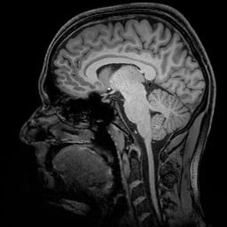
The human brain continuously collects information. However, we have only basic knowledge of how new experiences are converted into lasting memories. Now, an international team led by researchers of the University of Magdeburg and the German Center for Neurodegenerative Diseases (DZNE) has successfully determined the location, where memories are generated with a level of precision never achieved before. The team was able to pinpoint this location down to specific circuits of the human brain. To this end the scientists used a particularly accurate type of magnetic resonance imaging (MRI) technology. The researchers hope that the results and method of their study might be able to assist in acquiring a better understanding of the effects Alzheimer’s disease has on the brain. The science journal “Nature Communications” reports on their findings.
For the recall of experiences and facts, various parts of the brain have to work together. Much of this interdependence is still undetermined, however, it is known that memories are stored primarily in the cerebral cortex and that the control center that generates memory content and also retrieves it, is located in the brain’s interior. This happens in the hippocampus and in the adjacent entorhinal cortex. “It is been known for quite some time that these areas of the brain participate in the generation of memories. This is where information is collected and processed. Our study has refined our view of this situation,” explains Professor Emrah Düzel, site speaker of the DZNE in Magdeburg and director of the Institute of Cognitive Neurology and Dementia Research at the University of Magdeburg. “We have been able to locate the generation of human memories to certain neuronal layers within the hippocampus and the entorhinal cortex. We were able to determine which neuronal layer was active. This revealed if information was directed into the hippocampus or whether it traveled from the hippocampus into the cerebral cortex. Previously used MRI techniques were not precise enough to capture this directional information. Hence, this is the first time we have been able to show where in the brain the doorway to memory is located.”
For this study, the scientists examined the brains of persons who had volunteered to participate in a memory test. The researchers used a special type of magnetic resonance imaging technology called “7 Tesla ultra-high field MRI”. This enabled them to determine the activity of individual brain regions with unprecedented accuracy.
A Precision method for research on Alzheimer’s
“This measuring technique allows us to track the flow of information inside the brain and examine the areas that are involved in the processing of memories in great detail,” comments Düzel. “As a result, we hope to gain new insights into how memory impairments arise that are typical for Alzheimer’s. Concerning dementia, is the information still intact at the gateway to memory? Do troubles arise later on, when memories are processed? We hope to answer such questions.”
This work was supported by the Deutsche Forschungsgemeinschaft (DFG), SFB 779-TP A07.
Original publication
Laminar activity in the hippocampus and entorhinal cortex related to novelty and episodic encoding.
Anne Maass, Hartmut Schütze, Oliver Speck, Andrew Yonelinas, Claus Tempelmann, Hans-Jochen Heinze, David Berron, Arturo Cardenas-Blanco, Kay H. Brodersen, Klaas Enno Stephan, Emrah Düzel.
Nature Communications, 2014, doi: 10.1038/ncomms6547
If our reporting has informed or inspired you, please consider making a donation. Every contribution, no matter the size, empowers us to continue delivering accurate, engaging, and trustworthy science and medical news. Independent journalism requires time, effort, and resources—your support ensures we can keep uncovering the stories that matter most to you.
Join us in making knowledge accessible and impactful. Thank you for standing with us!

