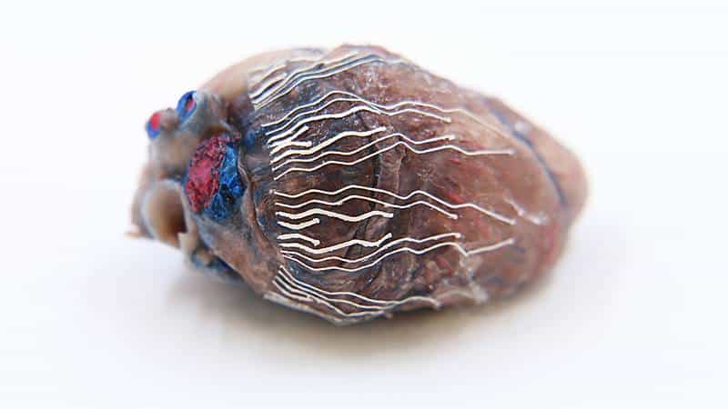
Surgeons may soon be able to localize critical regions in tissues and organs during a surgical operation thanks to a new, patent-pending Purdue University biosensor that can be printed in 3D using an automated printing system.
Chi Hwan Lee created the biosensor, which allows for simultaneous recording and imaging of tissues and organs during a surgical operation. Lee is the Leslie A. Geddes Assistant Professor of Biomedical Engineering in the Weldon School of Biomedical Engineering and assistant professor of mechanical engineering. Lee also has a courtesy appointment in materials engineering.
“Simultaneous recording and imaging could be useful during heart surgery in localizing critical regions and guiding surgical interventions such as a procedure for restoring normal heart rhythms,” Lee said.
Traditional methods to simultaneously record and image tissues and organs have proven difficult because other sensors used for recording typically interrupt the imaging process.
“To this end, we have developed an ultra-soft, thin and stretchable biosensor that is capable of seamlessly interfacing with the curvilinear surface of organs; for example the heart, even under large mechanical deformations, for example cardiac cycles,” Lee said. “This unique feature enables the simultaneous recording and imaging, which allows us to accurately indicate the origin of disease conditions: in this example, real-time observations on the propagation of myocardial infarction in 3D.”
By using soft bio-inks during the rapid prototyping of a custom-fit design, biosensors fit a variety of sizes and shapes of an organ. The bio-inks are softer than tissue, stretch without experiencing sensor degradation and have reliable natural adhesion to the wet surface of organs without needing additional adhesives. Kwan-Soo Lee’s research group in Los Alamos National Laboratory is responsible for the formulation and synthesis of the bio-inks.
A number of prototype biosensors using different shapes, sizes and configurations have been produced. Craig Goergen, the Leslie A. Geddes Associate Professor of Biomedical Engineering in Purdue’s Weldon School of Biomedical Engineering, and his laboratory group have tested the prototypes in mice and pigs in vivo.
“Professor Goergen and his team were successfully able to identify the exact location of myocardial infarctions over time using the prototype biosensors,” Lee said. “In addition to these tests, they also evaluated the biocompatibility and anti-biofouling properties of the biosensors, as well as the effects of the biosensors on cardiac function. They have shown no significant adverse effects.”
Research about the biosensor has been published in the peer-reviewed Nature Communications.
The Purdue Research Foundation Office of Technology Commercialization has filed a patent application on Lee’s biosensor. For licensing information, please contact Dipak Narula of OTC and reference track code 2021-LEE-69211. Other steps taken to develop the sensor include exploring further applications of the bio-inks into various printable biosensors with a tailored design to fit other organs such as the stomach, which requires even higher stretchability than the heart.
Writer: Steve Martin, [email protected]
Source: Chi Hwan Lee, [email protected]
ABSTRACT
Rapid Custom Prototyping of Soft Poroelastic Biosensor for Simultaneous Epicardial Recording and Imaging
Bongjoong Kim1, Arvin H. Soepriatna2, Woohyun Park1, Haesoo Moon2, Abigail Cox3, Jianchao Zhao4, Nevin S. Gupta4, Chi Hoon Park4,5, Kyunghun Kim2, Yale Jeon2,6, Hanmin Jang2,6, Dong Rip Kim6, Hyowon Lee2, Kwan-Soo Lee4,*, Craig J. Goergen2,*, Chi Hwan Lee1,2,7,*
1School of Mechanical Engineering, Purdue University, West Lafayette, IN 47907, USA. 2Weldon School of Biomedical Engineering, Purdue University, West Lafayette, IN 47907, USA. 3Department of Comparative Pathobiology, Purdue College of Veterinary Medicine, West Lafayette, IN, USA. 4Chemical Diagnostics and Engineering, Los Alamos National Laboratory, Los Alamos, New Mexico 87545, USA. 5Department of Energy Engineering, Gyeongnam National University of Science and Technology, Jinju-Si 660-758, Republic of Korea. 6School of Mechanical Engineering, Hanyang University, Seoul 04763, Republic of Korea. 7Department of Materials Engineering, Purdue University, West Lafayette, IN 47907, USA. †These authors contributed equally to this work. *Correspondence and requests for materials should be addressed to C.H.L. (email: [email protected]) or C.J.G. (email: [email protected]) or K.-S.L. (email: [email protected])
ABSTRACT
The growing need for the implementation of stretchable biosensors in the body has driven rapid prototyping schemes through the direct ink writing of multidimensional functional architectures. Recent approaches employ biocompatible inks that are dispensable through an automated nozzle injection system. However, their application in medical practices remains challenged in reliable recording due to their viscoelastic nature that yields mechanical and electrical hysteresis under periodic large strains. Herein, we report sponge-like poroelastic silicone composites adaptable for high-precision direct writing of custom-designed stretchable biosensors, which are soft and insensitive to strains. Their unique structural properties yield a robust coupling to living tissues, enabling high-fidelity recording of spatiotemporal electrophysiological activity and real-time ultrasound imaging for visual feedback. In vivo evaluations of custom-fit biosensors in a murine acute myocardial infarction model demonstrate a potential clinical utility in the simultaneous intraoperative recording and imaging on the epicardium, which may guide definitive surgical treatments.
If our reporting has informed or inspired you, please consider making a donation. Every contribution, no matter the size, empowers us to continue delivering accurate, engaging, and trustworthy science and medical news. Independent journalism requires time, effort, and resources—your support ensures we can keep uncovering the stories that matter most to you.
Join us in making knowledge accessible and impactful. Thank you for standing with us!

