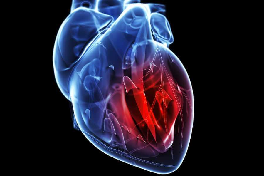
A new study reveals that the heart contains a complex network of neurons that forms its own “little brain,” capable of sophisticated information processing and even generating rhythmic activity independently of the brain.
Published in Nature Communications | Estimated reading time: 4 minutes
Scientists have long known that the heart houses its own network of neurons, called the intracardiac nervous system (IcNS). But this system’s true complexity has remained largely mysterious – until now. Using cutting-edge techniques including single-cell RNA sequencing and precise electrical recordings, researchers have created the first comprehensive map of the heart’s internal neural network in zebrafish, revealing an unexpectedly sophisticated system that shares striking similarities with the human heart.
The research team found that this neural network contains diverse types of neurons that use different chemical signals to communicate. While the majority of heart neurons (80.78%) were found to use acetylcholine, the researchers also identified neurons that use glutamate (8%), GABA (6%), and serotonin (5%), demonstrating a level of complexity previously unrecognized.
Most significantly, the study identified a subset of neurons that display pacemaker-like properties, similar to the neurons that generate rhythmic patterns in the central nervous system. These neurons exhibit specific characteristics including SAG potential and post-inhibitory rebound firing – properties associated with generating rhythmic activity. This suggests the heart’s neural network isn’t just relaying signals from the central nervous system, but actively processing and generating its own commands.
The researchers used zebrafish for their study because these animals’ hearts share key similarities with human hearts in terms of rate, cardiomyocyte action potential duration, and morphology. Using a sophisticated whole-mount ex-vivo preparation technique that allowed them to record electrical activity from individual heart neurons while keeping the heart intact, the team discovered four distinct types of neurons with different firing patterns: Single Spike type, Adaptive type, Repetitive type, and Bursting type.
This research supports a model suggesting the vertebrate heart has a two-layer localized control system: a nodal pacemaker responsible for generating the inherent heart rate, and a neuronal regulatory module that determines the operational range after integrating central and peripheral information. This detailed understanding of the heart’s neural system opens new avenues for investigating cardiac function and regulation.
Glossary
- Intracardiac Nervous System (IcNS): The network of neurons embedded within the heart’s wall that helps regulate cardiac function.
- Neurotransmitters: Chemical signals used by neurons to communicate, including acetylcholine, glutamate, GABA, and serotonin.
- Pacemaker Neurons: Specialized nerve cells capable of generating rhythmic patterns of activity.
- Single-cell RNA Sequencing: A technique that allows researchers to study the genetic activity of individual cells.
Test Your Knowledge
What is the primary neurotransmitter used by heart neurons?
Acetylcholine is the main neurotransmitter used by the majority (80.78%) of heart neurons.
How many distinct types of neurons did the researchers identify based on firing patterns?
The researchers identified four distinct types of neurons with different firing patterns: Single Spike, Adaptive, Repetitive, and Bursting types.
What key characteristics do the pacemaker-like neurons exhibit?
These neurons display SAG potential and post-inhibitory rebound firing, properties that are associated with generating rhythmic activity.
What are the two layers of the heart’s control system according to the research?
The vertebrate heart has a nodal pacemaker that generates the inherent heart rate, and a neuronal regulatory module that determines the operational range by integrating central and peripheral information.
Enjoy this story? Subscribe to our newsletter at scienceblog.substack.com.
If our reporting has informed or inspired you, please consider making a donation. Every contribution, no matter the size, empowers us to continue delivering accurate, engaging, and trustworthy science and medical news. Independent journalism requires time, effort, and resources—your support ensures we can keep uncovering the stories that matter most to you.
Join us in making knowledge accessible and impactful. Thank you for standing with us!

