Scientists have turned to cameras designed for quantum measurements to capture the earliest stages of life, marking a significant advancement in how researchers can observe developing embryos while minimizing potential damage.
Researchers at the University of Adelaide’s Centre of Light for Life have demonstrated for the first time that ultrasensitive cameras—including those capable of detecting individual packets of light at each pixel—can effectively image living embryos in their natural state with minimal disruption.
“Damage from illumination is a real concern which can often be overlooked. Using the lowest level of light possible, together with these very sensitive cameras is important for understanding biology in live and developing cells,” said Professor Kishan Dholakia, director of the Centre of Light for Life.
The research team tested this cutting-edge technology on developing embryos as part of a pre-clinical trial, with results published in the journal APL:Photonics. The project united specialists across multiple disciplines, including optics, biology, laser physics, and microscopy.
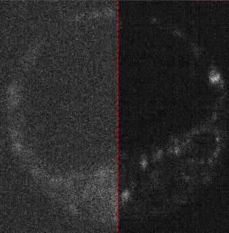
At the core of this innovation is the ability to detect extremely weak signals produced naturally within living cells when illuminated with minimal light—a capability that conventional imaging techniques struggle to achieve without potentially harmful higher light levels.
Lead author and PhD student Zane Peterkovic explained the technical challenge: “A lot of natural compounds in cells light up when illuminated, and this can tell us a lot about what we’re looking at, but unfortunately the signal is very weak.”
The advancement comes as fertility research increasingly seeks non-invasive methods to assess embryo health. Current clinical IVF practices often rely on visual assessment under microscopes to select viable embryos, but providing more detailed biochemical information without disturbing development could potentially improve success rates.
“These samples are living, developing specimens that serve as a foundation for studies supporting advancements in clinical IVF,” noted Professor Dholakia.
What makes this technology particularly intriguing is its roots in quantum physics. The cameras operate at such extreme sensitivity levels that quantum mechanics—typically associated with subatomic particles rather than medical imaging—becomes relevant to their operation.
“Digital camera technology has advanced to the point where fundamental physics concepts like quantum mechanics become important and relevant,” Peterkovic said.
The research went beyond simply attaching advanced cameras to microscopes. The team developed new methodologies to compare image quality across different cameras and even explored how artificial intelligence could enhance the results.
“We even explored how AI can be used to remove noise from the captured images, which is essentially static because the camera struggles to capture enough light,” said Peterkovic. “These steps go beyond just putting the camera in the microscope to take pictures.”
The latest generation of quantum cameras can count individual packets of light energy—called photons—at each pixel, representing the ultimate limit in gentle imaging. This extreme sensitivity allows researchers to observe cellular processes with minimal interference from the observation process itself—a concept reminiscent of the observer effect in quantum physics, where the act of measurement can alter what’s being measured.
Associate Professor Kylie Dunning, who leads the Reproductive Success Group with the Robinson Research Institute and was part of the research team, has previously investigated methods for improving embryo assessment through optical techniques.
The interdisciplinary approach highlights a growing trend in biological imaging, where techniques originally developed for physics experiments find new applications in life sciences. Similar technology crossovers have led to breakthroughs in brain imaging, cancer detection, and drug discovery.
Looking ahead, the researchers plan to extend their work into quantum imaging, where quantum states of light themselves may be used to gather even more information from biological samples without increasing light exposure.
This approach could eventually lead to more accurate techniques for assessing embryo viability, potentially improving IVF success rates by allowing embryologists to select the healthiest embryos with greater confidence while minimizing any potential light-induced damage during the assessment process.
The research was supported by funding from the Australian Research Council, highlighting the continued investment in cutting-edge optical technologies for biological applications.
As Professor Dholakia summarized: “Modern imaging technology is very exciting with what it enables us to see.”
If our reporting has informed or inspired you, please consider making a donation. Every contribution, no matter the size, empowers us to continue delivering accurate, engaging, and trustworthy science and medical news. Independent journalism requires time, effort, and resources—your support ensures we can keep uncovering the stories that matter most to you.
Join us in making knowledge accessible and impactful. Thank you for standing with us!