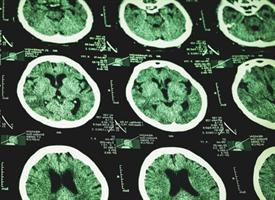
A new study out today in the Journal of Neuroscience shows that traumatic brain injury can disrupt the function of the brain’s waste removal system. When this occurs, toxic proteins may accumulate in the brain, setting the stage for the onset of neurodegenerative diseases such as Alzheimer’s and chronic traumatic encephalopathy.
“We know that traumatic brain injury early in life is a risk factor for the early development of dementia in the decades that follow,” said Maiken Nedergaard, M.D., D.M.Sc., co-director of the University of Rochester Center for Translational Neuromedicine and senior author of the article. “This study shows that these injuries set into motion a cascading series of events that impair the brain’s ability to clear waste, allowing proteins like tau to spread throughout the brain and eventually reach toxic levels.”
The findings are the latest in a series of new insights that are fundamentally changing the way scientists understand neurological disorders. These discoveries are possible due to a study published in 2012 in which Nedergaard and her colleagues described a previously unknown system of waste removal that is unique to the brain which researchers have dubbed the glymphatic system.
The brain is essentially closed off from the rest of the body by a complex system of molecular gateways, called the blood-brain barrier, that tightly control what enters and exits the brain. Consequently, the body’s normal waste removal system does not extend to the brain.
As with the rest of the body, the timely removal of waste from the brain is essential to prevent the unchecked accumulation of toxic proteins and other debris. However, until recently no one was entirely clear how the brain accomplished this.
Nedergaard and her colleagues showed that mice, whose brains are remarkably similar to humans, possess what amounts to a plumbing system that piggybacks on blood vessels to pump cerebral spinal fluid (CSF), the fluid surrounding the brain, through brain tissue, flushing away the waste from the spaces between the brain’s cells.
Recent studies have shown that the glymphatic system is more active during sleep, which may explain why sleep is so refreshing to the mind, and that its function declines with age.
“The failure of the glymphatic system may be one of the reasons that the aging brain is so vulnerable to diseases like Alzheimer’s,” said Jeffrey Iliff, Ph.D., co-author of study and an assistant professor at Oregon Health and Science University. “It’s striking that the same changes that we see in the aging brain are mirrored in the young brain after traumatic brain injury. It suggests that these events may be the common link to neurodegeneration, between what happens in the elderly and what happens after brain trauma.”
The new research focuses on the impact that traumatic brain injury has on the glymphatic system. It has been long observed that the protein tau plays an important role in the long-term damage sustained by the brain after a trauma. Tau helps stabilize the fibers, or axons, that nerve cells send out to communicate with their neighbors.
However, during trauma, large numbers of these proteins are shaken free from the axons to drift in the space between the brain’s cells. Once unmoored from nerve cells, these sticky proteins are attracted to each other and, over time, form increasingly larger “tangles” that can become toxic to brain function.
Under normal circumstances, the glymphatic system is able to clear stray tau from the brain. However, when the researchers studied the brains of mice with traumatic brain injury, they found that the trauma damaged the glymphatic system, specifically the ability of astrocytes – a support cell found in the brain – to regulate the cleaning process.
Astrocytes play a critical role in organizing the flow of CSF into the brain. Branches from the cells enclose the brain’s blood vessels creating a space– essentially a pipe within a pipe – into which CSF can follow the path of the blood vessels and flow into the interior of the brain. The branches of the astrocytes that form the outer “pipe” are lined with a massive number of structures known as water channels – or aquaporins – that help ensure the efficient flow of CSF along the blood vessels into and out of the brain. The researchers observed that after traumatic brain injury, the aquaporins lose their organization, impairing the flow of CSF into the brain.
“In order to clear waste the glymphatic system must pump CSF through the brain,” said Nedergaard. “This study would seem to indicate that the system is very delicate and that small changes in the organization of water channels can cause it to lose function.”
Long after the injury, the researchers noted that the excess tau was not being cleared from the animals’ brains and that tau had begun to aggregate throughout the brain. In animals with impaired aquaporin water channels, tau accumulated far more rapidly.
The researchers also had the animals perform a series of experiments to test their memory and cognitive abilities. The animals with traumatic brain injury all performed far worse than controls. Animals with impaired water channels function did even worse and showed no improvement over time.
“For a long time, we have viewed neurodegenerative diseases like Alzheimer’s as a supply problem, meaning that we believed the brain was producing too much tau or amyloid beta,” said Benjamin Plog, an M.D./Ph.D. student in Nedergaard’s lab and a co-author of the study. “It now appears that these conditions may ultimately be linked to a clearance problem, where something is preventing the glymphatic system from removing waste from the brain fast enough.”
Additional co-authors include Michael Chen, Lijun Yang, Itender Singh, and Rashid Deane with the University of Rochester, and Douglas Zeppenfeld with the Oregon Health and Science University. The study was supported with funding from the National Institute of Neurological Disorders and Stroke and the American Heart Association.
If our reporting has informed or inspired you, please consider making a donation. Every contribution, no matter the size, empowers us to continue delivering accurate, engaging, and trustworthy science and medical news. Independent journalism requires time, effort, and resources—your support ensures we can keep uncovering the stories that matter most to you.
Join us in making knowledge accessible and impactful. Thank you for standing with us!

