“Numerous other studies have shown that people with schizophrenia have significantly smaller volumes of brain tissue than healthy controls. However, for at least a third of patients we looked at, this was not the case at all—their brains were almost completely normal,” says principal investigator Christos Davatzikos, the Wallace T. Miller Professor of Radiology in the Perelman School of Medicine. “In the future, we’re not going to be saying, ‘This patient has schizophrenia,’ We’re going to be saying, ‘This patient has this subtype’ or ‘this abnormal pattern,’ rather than having a wide umbrella under which everyone is categorized.”
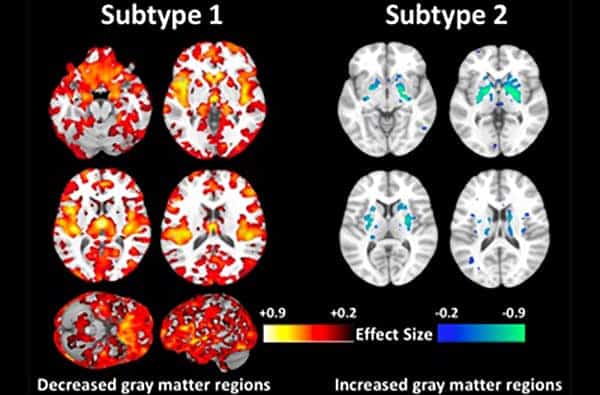
The researchers found that 115 patients with schizophrenia, or nearly 40 percent, did not have the typical pattern of reduced gray matter volume that has been historically linked to the disorder. In fact, their brains showed increases of brain volume in the middle of the brain, in an area called the striatum, which plays a role in voluntary movement. When controlling for differences in medication, age, and other demographics, the researchers could not find any clear explanation for the variation.
“The subtype 2 patients are very interesting, because they have similar demographic and clinical measures with subtype 1, and the only differences were their brain structures,” says Ganesh Chand, a lead author and postdoctoral researcher in the radiology department.
Read more at Penn Medicine News.
If our reporting has informed or inspired you, please consider making a donation. Every contribution, no matter the size, empowers us to continue delivering accurate, engaging, and trustworthy science and medical news. Independent journalism requires time, effort, and resources—your support ensures we can keep uncovering the stories that matter most to you.
Join us in making knowledge accessible and impactful. Thank you for standing with us!