

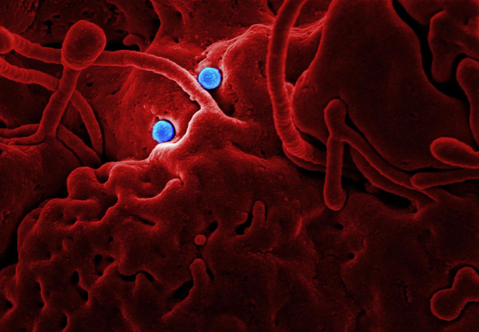
A team based at EMBL Hamburg has produced the first large-scale molecular map of how influenza A physically grabs hold of human proteins inside living, infected cells — and one of its most striking findings is that the virus systematically dismantles a set of tiny droplet-like structures in the cell nucleus called paraspeckles, apparently to free up proteins it needs for its own replication.
The work, published this week in Nature Microbiology, pairs a chemistry trick called cross-linking mass spectrometry with a modified version of the AlphaFold protein-structure algorithm. The combination let researchers catch viral and human proteins in the act of touching each other, then reconstruct what those handshakes look like in three dimensions.
It is, in effect, a wiring diagram for a hijacking.

Why mapping the flu at this resolution matters
Seasonal influenza still causes 3 to 5 million cases of severe illness worldwide each year and is linked to as many as 650,000 deaths annually, according to EMBL’s summary of the study. Influenza A, the subtype the team focused on, is the one that has driven every flu pandemic on record, from the 1918 outbreak to the H1N1 wave of 2009.
Understanding how the virus rewires a human cell is not academic. Every direct contact between a viral protein and a human protein is a potential drug target — a molecular seam where an antiviral could pry the machinery apart.
Until now, most maps of those contacts came from cells that had been broken open first. That approach captures interactions in a kind of biochemical soup, stripped of the compartments and gradients that make a real cell a real cell. The Hamburg team wanted to see the wiring while the lights were still on.
How the method works
Cross-linking mass spectrometry, or XL-MS, uses small chemical tethers that snap shut when two proteins are close enough to touch. Once the cell is later broken down for analysis, those tethers stay in place, marking which proteins were in contact and, crucially, which parts of them were touching.
Feed that positional data into a structure predictor and the abstract list of interactions turns into something closer to a 3D blueprint. The team used a customized version of AlphaFold, the protein-folding system whose original developers shared the 2024 Nobel Prize in Chemistry.
According to the EMBL announcement, Boris Bogdanow, a junior research group leader at the Institute of Virology at Charité, explained that the XL-MS technique captures protein-protein interactions in infected cells while providing structural information about these interactions.
The payoff is a dataset that describes not just which human proteins the virus grabs, but how it grabs them.
Hemagglutinin gets a chaperone escort
One half of the map traces the journey of hemagglutinin, the spiky protein that gives flu strains names like H1 or H5 and that the virus uses to latch onto human cells.
Hemagglutinin has to be folded, modified and shipped to the cell surface before new virus particles can bud off. The new work shows the virus co-opting the cell’s own protein-transport network to do exactly that — recruiting human chaperones and modifying enzymes at specific contact points along the way.
That handoff chain is the kind of detail that matters for antiviral design. Blocking a viral protein directly often selects for resistant mutants. Blocking the human protein the virus depends on is harder for the virus to evade, because it cannot easily mutate its host.
The same logic drives newer therapies for other viruses, including the class of herpes drugs that jam the viral replication engine by targeting host-virus interfaces.
The paraspeckle surprise
The second half of the map is the part that startled the researchers.
Paraspeckles are small, droplet-like compartments inside the cell nucleus. They are not membrane-bound organelles but condensates — protein-and-RNA droplets that form and dissolve depending on cellular conditions. They are thought to help regulate gene expression and to sequester certain RNA-binding proteins.
In every flu-infected cell line the team looked at, and across multiple flu strains, the paraspeckles dissolved.
Iuliia Kotova, the study’s first author and a former predoctoral fellow at EMBL Hamburg, noted that the behavior of paraspeckles was the most surprising finding. Kotova explained that observing the consistent dissolution of these organelles across all cell lines and flu strains tested suggested this was likely a deliberate viral strategy rather than merely a side effect of infection.
When the droplets fall apart, they release the RNA-binding proteins they were holding. Those proteins are exactly the kind of molecular tools influenza needs to copy its own RNA genome. Dissolving paraspeckles may also blunt the cell’s antiviral response, though the researchers frame that second effect more cautiously.
A snapshot, not a movie — yet
The current map captures a single moment in the infection cycle. Flu, though, is a fast-moving process: entry, uncoating, replication, assembly and budding all unfold within hours, and the protein interactions the virus depends on shift as it goes.
Jan Kosinski, a group leader at EMBL Hamburg, said the work offers a new approach to studying flu-host interactions within their natural cellular context while gaining structural insights. Kosinski noted that the current results represent a single moment during infection, suggesting future research could examine flu-host interactions throughout the complete infection cycle.
The next step is a time series — repeating the XL-MS mapping at successive points after infection to build something more like a stop-motion film of the takeover.
Why H5N1 is the elephant in the room
The method was demonstrated on seasonal influenza A strains grown in cell culture. But the approach is general, and the researchers explicitly flag H5N1 — the highly pathogenic avian flu now circulating in dairy cattle and wild birds — as an obvious next target.
H5N1’s toll on wildlife is already visible. Bass Rock, the world’s largest colony of northern gannets, saw its population fall by almost a third after the recent bird flu wave, and monitoring efforts along the Thames continue to track outbreaks in waterfowl, according to BBC coverage of ongoing swan surveys. Human cases have so far been rare, but every additional mammalian host is another opportunity for the virus to adapt.
Applying the Hamburg map to H5N1 would reveal whether the avian strain uses the same paraspeckle-dissolving trick and the same chaperone highways — and whether the same drug targets are on the table.
Implications for vaccines and antivirals
Current flu vaccines mostly train the immune system to recognize hemagglutinin and neuraminidase on the virus surface. That works, but only until the virus mutates those proteins enough to slip past. Broader-acting vaccines aim at conserved features that the virus cannot easily change — a strategy familiar from work on Ebola’s shape-shifting defenses.
The Hamburg map suggests another axis of attack. If a specific human chaperone is essential for hemagglutinin folding, a small molecule that briefly blocks that chaperone could stall flu replication regardless of strain. If paraspeckle dissolution is required for efficient viral RNA synthesis, drugs that stabilize those condensates might do the same.
Neither is a next-year proposition. Host-directed antivirals have to walk a narrow line between blocking a virus and disrupting normal cell function.
What the study delivers is the parts list. Roughly a century after the 1918 pandemic and two decades into the AlphaFold era, researchers can finally read the flu’s takeover instructions off the cell itself, one bond at a time.