Timely detection and accurate segmentation of acute ischemic stroke (AIS) lesions on magnetic resonance images (MRIs) are essential for the triaging patient for endovascular therapy. Lesion segmentation is a routine process where the abnormal areas within brain images are qualitatively and manually picked by expert radiologists. However, manual lesion segmentation is time consuming and suffers from operator-bias. Accordingly, efficient and low-cost approaches for AIS lesion screening are yet to be introduced.
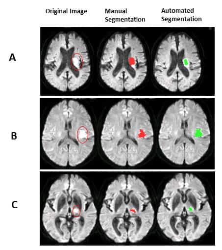
This research introduces a novel and fully automated technique for detection and segmentation of AIS lesions on MRIs and classification of images into stroke and none-stroke. This fully automated anomaly-detection method compares diffusion weighted images (DWIs) and apparent diffusion coefficients (ADC) images of the subjects with a group of healthy images in voxel-level. Areas with hyperintensity on DWI and hypointensity on ADC are identified as lesions and saved as lesion masks. The lesion segmentation method was investigated on approximately 100 cases. Since there is a risk of false lesion identification due to the artifacts, noises, and image low resolution, the lesion masks created by the method are screened and filtered via a binary classifier which either confirms that the created lesion mask contains a real AIS lesion or not. The classification performance was evaluated on about 200 MRIs.
The published results in the Journal of Neuroscience Methods show good agreement with the manually drawn lesions by experts (gold standard). The whole approach, including lesion segmentation and image classification, is straightforward, fast and does not require high computation power and memory.
“We believe that this method has the capacity to be implemented on an ordinary desktop workstation integrated into the routine clinical diagnostic pipelines of the hospitals. This approach can help the radiologists to speed up the workflow of lesion detection and to reduce the operator bias in lesion segmentation owing to the reproducibility of the method”, tells project researcher Sanaz Nazari-Farsani from Turku PET Centre.
If our reporting has informed or inspired you, please consider making a donation. Every contribution, no matter the size, empowers us to continue delivering accurate, engaging, and trustworthy science and medical news. Independent journalism requires time, effort, and resources—your support ensures we can keep uncovering the stories that matter most to you.
Join us in making knowledge accessible and impactful. Thank you for standing with us!