

The once-popular mitochondrial free radical theory of aging proved to be too glib. Aging isn’t fundamentally about dispersed damage; rather, dispersed damage is a result when the body’s defenses stand down in old age. Nevertheless, the mitochondria do play a role in aging, largely through signaling and apoptosis. Antioxidants targeted to mitochondria may be an exception to the rule that antioxidants don’t prolong lifespan. And other supplements and strategies that either promote production of new mitochondria or enhance their efficiency of operation show promise for modest lifespan extension.
Growing new mitochondria
A ketogenic diet leads to generation of new mitochondria, as do caloric restriction and exercise. Exercise when the body is starved for sugar (low glycogen) is the most potent stimulator of new mitochondrial growth. Exercise while fasting, or continue to exercise after you “hit your wall”.
Hormones that promote mitochondrial proliferation include thyroxin, estrogens, and glucocorticoids. Promoting new mitochondria has a tendency simultaneously to suppress apoptosis, programmed cell death [ref]. At later ages, apoptosis of cells that are still functional tends to be a larger problem than the failure of cancerous cells to eliminate themselves by apoptosis. In other words, suppressing apoptosis is (on balance) a good thing for anti-aging, but the downside is it can also increase risk of cancer.
Ubiquinone=CoQ10
Coenzyme Q-10 (aka ubiquinone) is an essential part of mitochondrial chemistry, shuttling electrons along their way to the ATP molecules that mitochondria generate as their primary energy export to the cell. It’s often called an antioxidant, but that’s not the primary role of CoQ10.
As a supplement, it is well-established with a good reputation. There is lots of evidence for benefits to health markers, especially athletic endurance, several aspects of heart health, and erectile dysfunction. If you have fibromyalgia or if you are taking statins, CoQ10 is strongly indicated. For chronic fatigue syndrome, it’s definitely worth trying.
But there’s no reason to expect it will increase your life expectancy. Supplementing with ubiquinone increases the lifespan of worms but not mice or rats [ref, ref].
Worms that cannot make unbiquinone live 10 times as long. Just saying…
A few years ago, ubiquinol was introduced as a more bioavailable form of ubiquinone. It’s more expensive, but there is not clear evidence that it is more bioavailable.
PQQ
Pyrroloquinoline quinone is helpful but not necessary part of mitochondrial chemistry. Bacteria make a lot of it; plants less; mammals only tiny quantities. Mice completely deprived of PQQ show growth deficiency, but the amount that they need is tiny compared to the quantities in PQQ supplements.
PQQ is a growth factor for bacteria, and the principal health claim for PQQ is that it can stimulate growth of new mitochondria. The evidence is based on biochemistry and cell cultures. In live mice, it has been shown that PQQ deficiency results in a mitochondria deficiency, but not that large quantities of PQQ lead to more mitochondria.
Bill Faloon (LEF) and Joseph Cohen (selfhacked) are big fans of PQQ, and you can read a list of benefits here. Cohen claims PQQ helps with sleep quality and nerve growth, leading to better cognitive function.
Small quantities of PQQ can be absorbed from many plant foods, but not animal foods. Much larger quantities come in supplement form. 100 g of tofu has just 2µg (micrograms). Supplements are usually 5-20 mg, hundreds of times as much as you’re likely to get from a vegetarian diet. Here is a table of PQQ concentrations in foods:
SkQ and MitoQ
These are two closely related molecules, originally synthesized in Russia in the 1970s, but it wasn’t until the 1990s that their therapeutic value was documented by two New Zealand scientists. One end of the molecule is CoQ10 (or a version found in plants, claimed to be even more powerful as an antioxidant). The other end of the molecule is an electric tugboat that pulls the molecule into mitochondria.
I’ve written a detailed report three years ago. At the time, I noted that the Russians claimed to extend lifespan of mice modestly with SkQ, and SkQ was found (also in Russian labs) to be a powerful rejuvenant for aging eyes. The Russians sell SkQ as eye drops. The Kiwis sell MitoQ as skin cream and also as pills.
Earlier this year, the Russian labs announced that SkQ had substantially extended lifespan of a mouse strain that was short-lived because of a mitochondrial defect. None of the Russian claims have been reproduced in Western labs. Three years ago, I was inclined to give the Russians the benefit of the doubt, but now I’m starting to wonder, since the New Zealand company has a laboratory arm, and they haven’t announced anything nearly so impressive.
Humanin and her sisters
Mitochondria have ringlets of their own DNA, encoding just 37 genes. (That doesn’t mean that the mitochondria only need 37 proteins; the great majority of proteins needed by mitochondria are coded in chromosomes of the cell nucleus, and transported to the mitochondria as needed.) Just 16 years ago, the first mitochondrial-coded protein to be discovered was named Humanin, because it was found to improve cognitive function to dementia patients, restoring some of their “humanity”. In addition to being neuroprotective, humanin promotes insulin sensitivity. Humannin’s action is not confined to the mitochondrion in which it was produced, but in fact it circulates in the blood as a signal molecule. Blood levels of humanin decline with age.
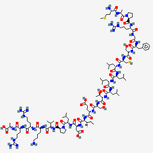
In experiments with mice, humanin injections have been shown to protect against disease. Lifespan assays with humanin are not yet available.
To date, HN and its analogs have been demonstrated to play a role in multiple diseases including type 2 diabetes (25, 43), cardiovascular disease (CVD) (2, 3, 47), memory loss (48), amyotrophic lateral sclerosis (ALS) (49), stroke (50), and inflammation (22, 51). The mechanisms that are common to many of these age-related diseases are oxidative stress (52) and mitochondrial dysfunction (53). Mitochondria are major source of ROS, excess of which can cause oxidative damage of cellular lipids, proteins, and DNA. The accumulation of oxidative damage will result in decline of mitochondrial function, which in turn leads to enhanced ROS production (53). This vicious cycle can play a role in cellular damage, apoptosis, and cellular senescence – contributing to aging and age-related diseases. Indeed, oxidative stress is tightly linked to multiple human diseases such as Parkinson’s disease (PD) (54), AD (55), atherosclerosis (56), heart failure (57), myocardial infarction (58), chronic inflammation (59), kidney disease (60), stroke (61), cancers (62, 63), and many types of metabolic disorders (64, 65). We and others have shown that HN plays critical roles in reducing oxidative stress (66–68). [2014 review]
Pinchas Cohen, MD (Dean, School of Gerontology, University of Southern California Davis, Los Angeles, California) is an expert in humanin, a protein (peptide) produced in mitochondria. Mitochondria are energy-generating organelles in cells, which have their own DNA separate from the DNA in the nucleus. The amount of DNA found in the mitochondria is much less than that found in the nucleus. As such, mitochondrial DNA contains codes for only a few proteins, humanin being one of them. Humanin was discovered by a search for factors helping to keep neurons alive in undiseased portions of the brains of Alzheimer’s disease patients. Humanin protects neurons against cell death in Alzheimer’s disease, as well as protecting against toxic chemicals and prions (toxic proteins)[ref]. Dr. Cohen’s team has shown that humanin also protects cells lining blood vessel walls, preventing atherosclerosis. In particular, they have shown that low levels of humanin in the bloodstream are associated with endothelial dysfunction of the coronary arteries (arteries of the heart).[ref] Humanin has also been shown to promote insulin sensitivity, the responsiveness of tissues to insulin. Because humanin levels decline with age, it is believed that reduced humanin contributes to the development of aging-associated diseases, including Alzheimer’s disease and type II diabetes. [Ben Best]
Personal notes: This lab near where I am visiting in Beijing is taking leadership in characterizing a group of short peptides similar in origin to humanin, and this company across the street from us is selling mitochondrial peptides.
If humanin were a patentable drug, there would be much excitement and multiple clinical trials for AD, probably leading to expansion into general anti-aging effects.
MOTS-c
This is another short peptide of mitochondrial origin, only recently discovered and characterized. I was alerted to its existence by a study from a USC lab that was written up here in ScienceBlog just this month (reprinted from a USC press release). Results are new but impressive. Mice injected with MOTS-c had more muscle mass, less fat, more strength and endurance. MOTS-c protected their insulin sensitivity when mice were fed a high fat diet [ref]. Lifespan studies haven’t been done yet.
Like humanin, MOTS-c is manufactured inside mitochondria from a template in mitochondrial DNA, but it is exported from the cell and appears in the bloodstream as a signal molecule. Blood levels of MOTS-c decline with age. It is a mini protein molecule with 16 amino acids, too big to survive digestion so it can’t be taken orally.
“MOTS-c holds much potential as a target to treat metabolic syndromes by regulating muscle and fat physiology, and perhaps even extend our healthy lifespan.”[ref]
Let’s keep your eyes on this one over the next year or two.
Gutathione / NAC
I’ve never heard anyone say a bad word about glutathione. It’s the antioxidant with no downside. Genetic modifications that upregulate glutathione have increased lifespan in worms, flies and mice.
For a long while, it has been assumed that you can’t eat glutathione, because it doesn’t survive digestion. Some researchers at Penn State disagree, finding impressive increases in tissue and blood levels when people were supplemented with up to 1 g per day raw glutathione. Liposomal glutathione is an oral delivery form that gets around the digestion problem, especially when taken with methyl donors like SAMe.
The herb Sylimarin=milk thistle may increase glutathione. For now, the precursor molecule N-Acetyl Cysteine (NAC) is the best-established supplement we have to promote glutathione. In the one available study, supplementing with NAC greatly increased lifespan in male but not female mice. NAC also increases lifespan in worms and flies.
-N-Acetylcysteine_Structural_Formulae.png)
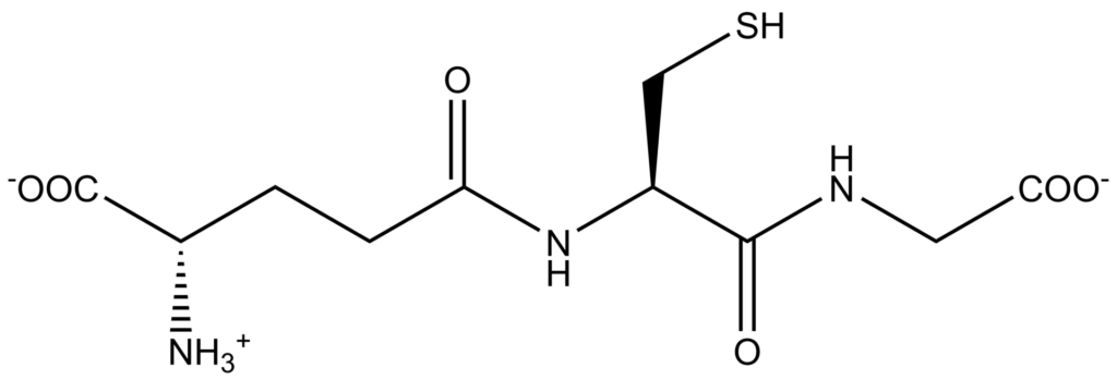
For the future, we might hope to do better. Less than 20% of the cell’s glutathione actually makes its way to the mitochondria, where it is most needed. There are esters of glutathione that, in theory, ought to be attracted into the mitochondria. They have been tested in cell culture only, but are more than ripe for animal testing [ref].
Nicotinamide Riboside (NR) and other NAD+ enhancers
The chemicals NAD+ and NADH are alternative, cycled forms of an intermediate in the process by which mitochondria make energy. Levels of NAD+/NADH decline with age. NR is a precursor to NAD+, and it has been demonstrated (preliminary results in humans) that NR supplementation increases blood levels of NAD+.
It may be awhile before we know for sure whether this leads to better health or longer lifespan. Niagen and Basis are heavily promoted with credible scientifists behind their products, and many early adopters offer subjective reports of short-term benefits. There is one mouse study claiming to pull a 3% extension of lifespan out of the noise, and perhaps I am less open to the finding because the article, published prominently in Science, seems so breathless in describing benefits.
Melatonin
The primary role of melatonin is to regulate the body’s sleep/wake cycle. Melatonin declines with age and the timing of our daily melatonin surge gets fuzzier and less reliable with age.sleep quality deteriorates. Sleep quality suffers.
Melatonin is well-established in mice as a modest longevity aid, although results have been inconsistent. 12 out of 20 studies showed a lifespan increase, and the remaining 8 showed no increase or decrease. Whether nightly supplementation affects mortality rates in humans has never been determined.
Melatonin is concentrated in mitochondria as much as 100-fold, and it may even be created there [ref], independent of the circulating melatonin that is secreted from the pineal gland at night. One of its actions is as a mitochondrial antioxidant and scavenger of ROS.
Twenty years ago, Walter Pierpaoli promoted melatonin as a sleep aid, cancer fighting hormone that would enhance your mood and your sex life while keeping you young. Russian labs have also been optimistic. My take is that melatonin is a legitimate anti-aging hormone, and is especially useful for those of us whose sleep is disrupted with age. It is widely available, cheap and safe. Unless you’re fighting jet lag, 1 to 2 mg at night is all you need.
Also worth mentioning
Magnesium is required for manufacture of glutathione. Selenium works along with glutathione. Omega-3 fatty acids can promote synthesis of glutathione. Acetyl L-carnitine transports fat fuels through the mitochondrial membrane. Alpha-lipoic acid is part of the mitochondrial energy metabolism.
The Bottom Line
Commercial interests can make some messages louder than others, and the health news we hear is affected by what is profitable as much as by what is healthy. Exercise is primary, but has no sales value. Of the supplements reviewed here, NAC is the best-established for mitochondrial health and a possible effect on lifespan. It is cheap and available. Liposomal glutathione is certainly more expensive and possibly more effective. Melatonin is even cheaper, and has been found to increase lifespan in multiple rodent studies, with broad benefits apart from modification of mitochondrial function. Humanin and MOTS-c, not yet close to commercial availability, seem to be promising substances to explore for health, though not for profits.
Discussion
64 reader comments
Imported threads are marked Archive. New comments are welcome and moderated for spam.
Josh -
I'm wondering if you've had any further contact with Dr. Skulachev and if he has any updates on when he expects SKQ1 to be approved in tablet form in the Russian Federation?
Is anyone familiar with the new startup that David Sinclair attached himself to named Cohbar? They are a public-traded biotech that claims to have a pipeline of many mitochondrial peptides. Does anyone know whether there is any animal research on Cohbar's peptides? It feels in their presentations like they are selling generalizations but I don't find any evidence that any animal studies exist. What reason is there to believe their peptides treat the diseases they want to treat?
Josh, as for SKQ1 I think you refer to this study on mutant mice.
https://peachpuff-wolverine-566518.hostingersite.com/492213/scientists-slow-aging-artificial-antioxidant-skq1/
The credibility of this study is greatly enhanced by the cooperation with the Wenner Gren Institute in Stockholm.
Very interesting. The Aging paper can be found here:
http://www.aging-us.com/article/101174/text
Looks like they have well documented lots of beneficial effects associated with SKQ1 and the life span curve is quite convincing. However, since they have used mtDNA mutator mice, I am not sure what to conclude. I hope we will see soon a life span study on normal mice with similar benefits and life extension.
Hi PeterB,
Thanks for posting very interesting paper.
Mitochondria certainly one of great forces in aging.
The paper post by Mark, "The path from mitochondrial ROS to aging runs through the mitochondrial permeability transition pore", Rottenberg, July 2017 is a really great paper which brings together 60 years of developing theory about ROS and mitochondria. I urge everyone to study this paper. I see mTOR and mitochondria as two great mountains in aging. We can now control mTOR so when have even better drugs for mitochondria, people may reach 105 and still be in good health.
Hi Dr. Green,
Thanks for pointing out this interesting paper. I guess the next step would be to find a compound that specifically inhibit the mPTP and to show that it extends the life span of normal mice. Such a compound would be expected to have a synergistic effect with Rapamycin, right?
Hi Aldebaran,
Thanks for your comment.
The idea of this paper is very large number of things are acting on mitochondria and action goes through the final common pathway of mPTP opening. A few specific things they mention are caloric restriction, physical activity and metformin through increase amount of NADH acting on mPTP. They also mention sirt3 which studies show is increase by angiotensin blockers and have major effect on mitochondria.
As regards synergistic effect, I regard rapamycin as acting on mTOR and metformin as acting on mitochondria. Note that metformin didn't have much effect on mouse lifespan alone; but when combined with rapamycin had a much bigger effect than rapamycin alone.
In studies, angiotensin blockers increased lifespan in normal mice 20% and that appeared to be mitochondria effect mediated by sirt3 and decrease superoxide all substances mentioned in this paper.
So I think we do have very many drugs acting on mitochondria and mPTP, it is just fantastically complicated. There is an entire world of mitochondria chemistry to be unraveled; but this paper posted by Mark is a great start.
Anti-aging medicine got a million-to-one lucky break with the discovery of a bacteria on Easter island that made rapamycin, a magic bullet for mTOR.
Doesn't look like any single magic bullet for mitochondria; but might take a dozen things combined.
My guess, if dying of aging at age 75, you are dying from elevated mTOR and if dying of aging at 100, you are dying from mitochondrial disease.
Anyone knowledgeable in mitochondria want to comment on the influence of the mitochondrial permeability transition pore in aging?
http://onlinelibrary.wiley.com/doi/10.1111/acel.12650/full
Seems a strong contender to me for the influence of mitochondria on maximum lifespan.
Thanks Mark.
This looks like one fantastic paper you posted which will need a very lot of study. I divide aging into early aging (65-90, the mTOR driven diseases) and late aging, the real inexorable killer. Mitochondria seems at the the center of late aging (what you called maximal lifespan) and this paper seems to zero in on what is going bad with mitochondria. While early aging is now well understood and can be controlled, late aging (what Blagosklonny called post aging syndrome) is one complete mystery.
Thanks Mark also,
It is also illustrative this work on rapamycin and spermidine and Autophagy in stem cell aging of 7 August http://onlinelibrary.wiley.com/doi/10.1111/acel.12655/full
Mitochondria are destroyed by ROS (which Denham Harman discovered in 1961) and no one will change it. The primary sector where this knowledge was used was food industry. Shortly after the discovery, "aging accelerating foods by ROS" began to be promoted.
I estimate that "accelerating aging" foods are able to reduce life by 20% on average. These foods "eats" about 50% of the population. Can you imagine how much less is spent on pensions when the average life span is reduced by 10%? Even a car dealer can not afford to spend so much on advertising.
By excluding these substances from your diet, you will prolong your life more than using methylene blue, rapamycin, and metformin altogether.
This paper is very supportive of TOR driven aging (classed here as metabolic rate) and the stability of mitochondrial DNA being the two largest factors on the difference in lifespans between mammals. Together they account for almost 80% of the variation!
https://www.ncbi.nlm.nih.gov/pubmed/18442324
If we believe what this paper is saying, and although I'm no statistics expert I believe we should, then we have to look to these two main drivers of aging before anything else.
For me, this paper posted by Mark is most important paper I have seen in 2017 and gives me very different view of mitochondria. Previously I had seen loss of mitochondria as just loss of a vital structure. Now I see mitochondria as potential 2 micron potential Chernobles. Through the mitochondrial permeability transition pore mitochondria can damage rest of cell setting off a downward spiral resulting in cell death.
Previously has seen this phenomenon in Parkinson's disease in which failure to have proper housekeeping and quality control as regards mitochondria leads to loss of mitochondria and death of neurons; but in Parkinson's disease limited to neurons in substantia nigra.
Looks like through mitochondrial permeability transition pore the process of cell damage from failing mitochondria can be a generalized phenomenon when have failure to timely remove defective mitochondria by normal process of mitophagy.
Insulin sensitivity tends to decline with age. Perhaps this might be one way to combat this decline?
http://www.medscape.com/viewarticle/841119_4
Intranasal Insulin Enhanced Resting-state Functional Connectivity of Hippocampal Regions in Type 2 Diabetes
Discussion
This study demonstrated that in diabetic and age-matched healthy subjects, intranasal administration of a single dose of insulin acutely increased resting-state functional connectivity between the hippocampal regions and multiple regions within the DMN (i.e., MFC, IPC, ACC, and PCC) that are linked to integrative higher cognitive functions. After placebo administration, connectivity between hippocampal regions and these DMN regions was lower in diabetic subjects as compared with healthy control subjects in several brain regions. After insulin administration, the cluster size differences between the diabetes and the control groups decreased by 44% in the MFC and by 95% in the ACC. After administration of intranasal insulin, the differences in functional connectivity between the diabetes and control groups were no longer significant.
These findings suggest that acute administration of insulin via intranasal delivery route may improve functional connections between brain regions involved in memory and cognitive processing in other domains.
The insulin resistance syndrome is associated with reduced brain insulin levels and sensitivity in age-related memory impairment and AD.[5,27–29] Brain insulin plays an important role as a neuromodulator in cognition,[4,5] energy homeostasis, food intake, sympathetic activity, neuron-astrocyte signaling, synapse formation, and neuronal survival.[7,30] Insulin has been shown to reinforce signaling in the dopamine-mediated brain reward system and modulate food intake and responses to reward stimuli.[31–33] Intranasal insulin increases rapidly in cerebrospinal fluid and binds to insulin receptors[34,35] in the olfactory bulb, several regions in the cerebral cortex including the autonomic network (e.g., insular cortex, dorsal root ganglia, nigro-striatal neurons), cerebellum,[36–38] hypothalamus, and hippocampus.[34,35,39]
T2DM is associated with impairment of hippocampus-dependent memory, and these effects are proportional to diabetes severity.[2] Resting-state functional connectivity is also altered in T2DM subjects, and the severity of impairment correlates with the degree of insulin resistance.[18,19] The effects of intranasal insulin on resting-state connectivity have not been studied. Diabetic subjects had worse baseline cognitive performance, especially in the memory and executive function domains. We have previously shown, in this cohort, that intranasal insulin may acutely improve visuospatial memory in older diabetic and healthy adults, and that this improvement of memory and verbal learning may be dependent upon vasodilation response in the middle cerebral artery territory and in particular insular cortex.[10] In diabetic subjects on insulin, better performance on the verbal fluency naming task was associated with stronger coefficient of connectivity between the right hippocampal region and ACC and lesser connectivity between the left hippocampal regions and the MFC for a more difficult category switching task. In control subjects on insulin, better performance on the visuospatial memory task (BVMT-R) tended to correlate with stronger connectivity between the left hippocampal region and PCC. Differences in relationships between cognition and connectivity between the right and left hippocampal regions are intriguing and reflect a complexity of the large-scale verbal fluency network that comprises of verbal fluency and orthographic discrimination subnetworks.[40] Set switching is a complex operation involving a number of different brain structures that usually include various parts of the dorsolateral and dorsomedial prefrontal cortex, as well as temporal regions where hippocampus is located.[41] Functional integration within the verbal fluency network declines with age and task difficulty. Low productive-difficult tasks are associated with significant decreases in connectivity. Therefore, the decreased connectivity between the left hippocampus and DMN regions may reflect inhibition of the left hippocampus as a result of the complex category switching process.[42] After placebo administration, we have observed a "deactivation pattern"[15,16] that is characterized by task-related decreases in activity and connectivity among several DMN regions. In other words, during a task, a better task-related performance is associated with a decrease in functional connectivity within DMN.
Looks like mitochondria can be transplanted into humans. Could this provide a way to restore mitochondria fitness in late life?
Mitochondrial transplantation: From animal models to clinical use in humans
http://www.sciencedirect.com/science/article/pii/S1567724917300053
There is no doubt mitochondria function less well with age - the question is why: Is it some in-built mechanism inherent to mitos themselves, or is it something regulated by the cell?
My money is on the second option. There's just no reason for these organelles to age outside of an external signal.
They are still a valid intervention point, but if I am correct healthy mitos will translate into a healthier, more vital person - just like being fit, but will not translate to longer life.
The way I view aging is the same way I view the universe of Infectious disease; a very large number of different diseases, malaria, tuberculosis, bubonic plague, HIV, etc.
We are now beginning to understand different diseases that make up aging. Elevated TOR driven diseases seems to be most important among the common age-related diseases and conditions in the 60-95 age group and the one cause can now effectively treat. However, mitochondrial diseases discussed in this section by Josh also seem very important.
This report on stem cells related to hypothalamus can be another age-related disease. The nature of disease described in this paper appears to be that stem cells from hypothalamus make a substance that circulates in CSF and promotes nerve cell health and this declines with age and loss of this substance produced by stems cells promotes certain changes associated with aging. If true, it is another newly discovered aging disease, which joins a growing list. However, as in the world of Infectious disease, each new infectious disease just adds to the list, it doesn't erase other diseases or become the Master of Infection.
Alan Green, M.D.
Having searched a while and found the New Scientist article again, I will post a copy here in case it disappears again. Your post on how the blood of young mice had a mysterious 'youthing' effect on old mice is hereby explained.
Stem cells in the brain’s hypothalamus help mice stay young
By Jessica Hamzelou
YOUR brain may be to blame for your ageing body. A small cluster of stem cells in the brain seems to help mice stay young, and injecting extra stem cells helps them live longer. One day anti-ageing drugs might be able to replicate the effect in people.
Ageing is a complicated process, involving DNA damage, chronic inflammation, and worn-out cells, but we don’t yet know which of these has the biggest impact on ageing. Dongsheng Cai at the Albert Einstein College of Medicine in New York has been investigating the role of the brain in ageing, since it controls most of our bodily functions.
His team previously found that the hypothalamus, which releases hormones that affect other organs, seems to affect how mice age. By interfering with a molecular pathway in the hypothalamus, the team extended the lifespan of mice by 20 per cent.
Cai’s team wondered whether stem cells here might influence ageing. Although stem cells in the hypothalamus create new neurons throughout life, the team noticed that mice start losing them in middle age – about 10 or 11 months old. By the time mice are 2 years old – around 70 in human years – the cells are basically all gone, says Cai.
Mice age faster if these stem cells are destroyed. “There was a decline in learning and memory, coordination, muscle mass, endurance and skin thickness,” says Cai. The mice died a few months earlier than untreated animals.
But injecting the hypothalamus with extra stem cells, taken from the brains of newborn mice, slowed down this premature ageing, and gave mice an extra two to four months of life (Nature, DOI: 10.1038/nature23282).
First the team had to modify the stem cells so that they kick-started an anti-inflammatory pathway in the mice, otherwise the cells died and the injections didn’t work. This suggests that it may be inflammation that usually causes the death of stem cells in the brain as we age.
The team found that the injected stem cells secreted a particularly large amount of microRNAs. These are small molecules that can affect the way genes work, and the types of microRNA in our blood are known to vary according to age. Cai isn’t sure how the stem cell microRNAs might be working, but they seem to reduce biological stress and inflammation, he says.
Cai thinks his team’s findings could one day lead to a treatment for ageing. Once the microRNAs have been identified, it might be possible to develop drugs that mimic their effects, he says.
This may have the potential to become a therapy in about 30 years, says Richard Faragher at the University of Brighton, UK, who says other teams are already working towards microRNA drug treatments. An alternative strategy would be to target inflammation more generally. “I can see us taking multiple approaches,” says Faragher.
This article appeared in print under the headline “Stem cell boost slows down ageing”
Thank you! Many people sent me links to the original Nature paper yesterday as it came out. I'm excited, too, and I'll write about it as soon as I have time to digest it.
- JJM
This story was also reported here Josh
https://theconversation.com/scientists-discover-how-the-brains-hypothalamus-controls-ageing-and-manage-to-slow-it-down-81510
Josh,
Did you see this?? It appears that scientists have stalled aging in mice if they injected new stem cells into the hypothalamus. The scientists reason that aging is caused by the hypothalamus, so if you keep hypothalamic cells young, you'll stall aging. This paper does support your hypothesis that aging is controlled by an internal clock.
https://www.nature.com/nature/journal/vaop/ncurrent/full/nature23282.html
Josh I posted a comment about this just published research on the impact of these snps on aging yesterday. But my comment has disappeared. So too has the link I provided to the New Scientist article on the same subject.
Both gone ? That's very odd.
However as these miRNA's circulate in the blood surely they are the aging signals you discussed last year in a post here. Or rather the lack of miRNA's in the blood are the aging program signal
Hi Josh, very good work! one note,
this article was published yesterday on the subject -07/24/2017,
I think it's worth reading for its depth, thanks for all
Review Article: Pro- and Antioxidant Functions of the Peroxisome-Mitochondria Connection and Its Impact on Aging and Disease
https://www.hindawi.com/journals/omcl/2017/9860841/
You referenced the work of several Russian scientists; I am reminded of they also identified the peptide - Epitalon - which acts on the Pineal gland and produces melatonin. Have you developed any opinions about the use of Epitalon for melatonin production.
I as disappointed that the Russian work has not been investigated by Western scientists; is this just "not invented here" syndrone.