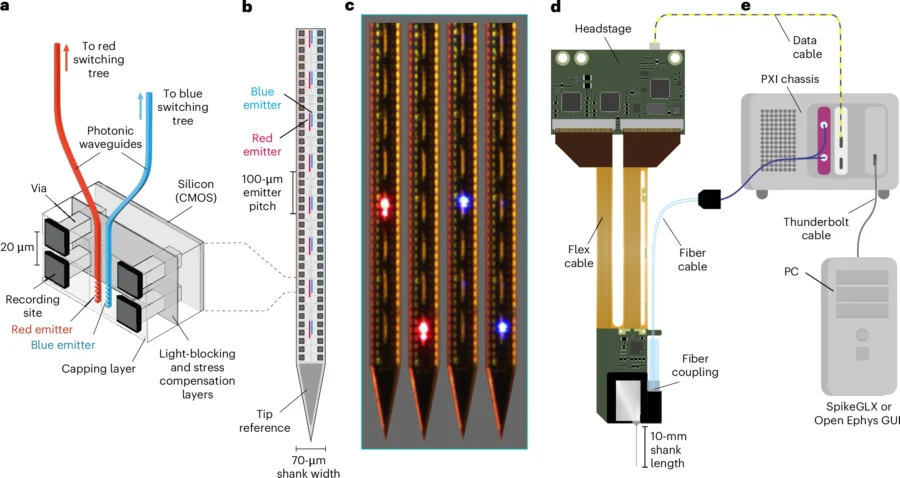
The thing is narrower than a single human hair, and along its silicon length sit close to a thousand tiny recording points and twenty-eight microscopic windows that spit out light. Slide it into the brain of a mouse and it does two jobs that neuroscientists have, for years, been forced to do separately. It eavesdrops on hundreds of neurons firing. And it tells particular ones, hand-picked by the experimenter, to switch on or shut up. Both at once, in the same animal, in the same instant.
That combination is the whole point of Neuropixels Opto, a device described in Nature Methods by an international team led from University College London and the Allen Institute in Seattle. It sounds like a small thing, sticking two existing tricks onto one sliver of silicon. It really isn’t.
For roughly two decades the field has leaned on two tools that don’t get along. Electrophysiology, the old workhorse, reads the electrical chatter of cells through electrodes. Optogenetics, the newer arrival, engineers neurons to respond to light so you can drive them like switches. Pair them and you can do something properly powerful: poke a specific population and watch, in real time, what the rest of the network does about it. The snag has always been the light. Brain tissue scatters and swallows it, so getting enough light deep into the brain usually meant shoving in a second device, an optical fibre or an array of miniature LEDs, which then muddied the very recordings you were trying to make.
Professor Matteo Carandini at UCL, one of the senior authors, puts the old problem plainly. “Combining the two has proved challenging, particularly in deeper brain regions, where delivering light without disrupting sensitive recordings is technically difficult,” he says.
Light that stays out of its own way
The fix is a clever bit of plumbing. Rather than generating light inside the brain (which is where the heat trouble starts), the team make it outside, with lasers, and pipe it down the shank through on-chip waveguides, essentially microscopic light pipes etched into silicon nitride. The emitters then fling the light sideways, away from the recording sites. That last detail matters more than it might seem. Point a bright pulse straight at a recording electrode and you get a photoelectric artefact that can top a millivolt, swamping the neural signal; aim it away and the artefact drops to around 30 microvolts, small enough to scrub out with routine processing.
Why bother with two colours, blue and red? Because it lets you talk to two genetically defined groups of cells in the same experiment, since the light-sensitive proteins each population carries respond to different wavelengths. The red, set at 638 nanometres, was deliberately tuned to dodge the wavelengths that blood absorbs most greedily, so it reaches further into tissue.
The packing is what’s genuinely hard to fabricate. The shank crams 960 recording sites and 2 sets of 14 emitters onto a strip 70 micrometres wide, and getting the photonics to sit on top of the recording circuitry without bending the shank or leaking stray light into the light-sensitive electronics took some doing. The bill, in manufacturing terms, is steep: the prototype needs around 740 processing steps to make, almost twice the roughly 400 that the standard Neuropixels probes require. Light is routed to any chosen emitter through a tree of tiny optical switches. And here the device showed its rough edges, the blue channel proved a bit temperamental, with some light leaking from emitters it wasn’t supposed to, so for the high-precision work the researchers leaned on red.
The cortex was less of a crowd than expected
Then came the part that surprised them. Co-lead author Dr Karolina Socha, a research fellow at the UCL Institute of Ophthalmology, has been using the probes to prod the cerebral cortex, the brain’s outer sheet that handles much of its heavy lifting. “We were surprised to discover that the activity of neurons in the cortex can be remarkably localised,” she says.
That cuts against expectation. The cortex is famously a tangle, its layers wired densely into one another, and the assumption had been that you couldn’t nudge a handful of neurons without setting off a crowd. When the team fired a single emitter, though, the cells that lit up sat in a tight band at the matching depth, roughly 150 micrometres of vertical spread, rather than spilling across the whole column.
Socha frames the shift in thinking carefully. Up to now, she says, the working assumption was that neurons are so interconnected there would be no way to switch some on without dragging many others along. The probes suggest otherwise: that these cells can act in concert and yet also, to a degree, go their own way.
The probes can do more than tickle cells, mind. In another set of experiments the team used them to drive local circuits, switching on inhibitory neurons and then watching those neurons clamp down on their neighbours through real synaptic connections, the kind of cause-and-effect loop that’s the bread and butter of how circuits actually compute. And in deeper structures like the striatum, the device proved handy for optotagging, flagging which recorded cells belong to which genetic type by seeing which ones answer the light. In one striatal recording it identified 25 of 39 cells; across the whole study, 261 in all.
This is where the medical hope creeps in, though it’s worth keeping it at arm’s length for now. Many disorders, schizophrenia, Alzheimer’s, Parkinson’s among them, involve neurons miscommunicating. A tool that can map healthy and faulty circuits at the level of individual cells, the UCL team suggest, might eventually point the way to more targeted treatments. Eventually being the operative word: this is mouse work, and a prototype at that.
What happens next is, in a sense, the least glamorous and most important bit. The team plan to do for these probes what they did for earlier Neuropixels, harden the fussy blue-light components, add detectors to monitor each emitter, shrink the whole package, and then manufacture the things in bulk and sell them at cost to labs around the world. Whether the surprising independence of those cortical neurons holds up elsewhere in the brain, nobody yet knows. But for the first time researchers have a single instrument that can both ask the question and listen for the answer.
DOI / Source: 10.1038/s41592-026-03076-z
Frequently Asked Questions
Why does it matter that one device can both record and control neurons at the same time?
Because it lets researchers test cause and effect directly rather than guessing at it. By switching specific neurons on or off and watching how the surrounding network responds in the same instant, scientists can establish which cells actually drive a given pattern of activity. That kind of causal evidence is far harder to get when recording and stimulation happen on separate devices or in separate animals.
How does the probe get light deep into the brain without cooking the tissue?
Instead of generating light inside the brain with miniature LEDs, which are inefficient and warm the tissue, the system makes light with external lasers and pipes it down the shank through tiny waveguides etched into silicon. The emitters then direct the light sideways, away from the recording electrodes, which also keeps electrical interference to a minimum. It is the routing, more than the light source, that makes the trick work.
Is it true that neurons in the cortex fire more independently than expected?
That is what these experiments suggest, at least in mice. The cortex is densely interconnected, so the assumption had been that stimulating a few neurons would inevitably pull many others along. When the team activated a single emitter, though, the responding cells clustered in a tight band rather than spreading through the whole column, hinting that local populations can act with more autonomy than the wiring implies.
Could this lead to new treatments for conditions like Alzheimer’s or schizophrenia?
Possibly, but not soon. Many neurological and psychiatric disorders involve neurons communicating abnormally, and a tool that maps circuits cell by cell could help reveal what goes wrong. For now this is prototype technology tested in mice, so any clinical payoff is a long way down the road.
What is still holding the technology back?
Two things, mainly. The blue-light channel proved unstable at high intensity, leaking light from the wrong emitters, so the researchers relied on red light for precision work. And the probes are demanding to manufacture, needing nearly twice as many fabrication steps as standard Neuropixels, which the team will have to streamline before producing them at scale.
Discover more from NeuroEdge
Subscribe to get the latest posts sent to your email.