

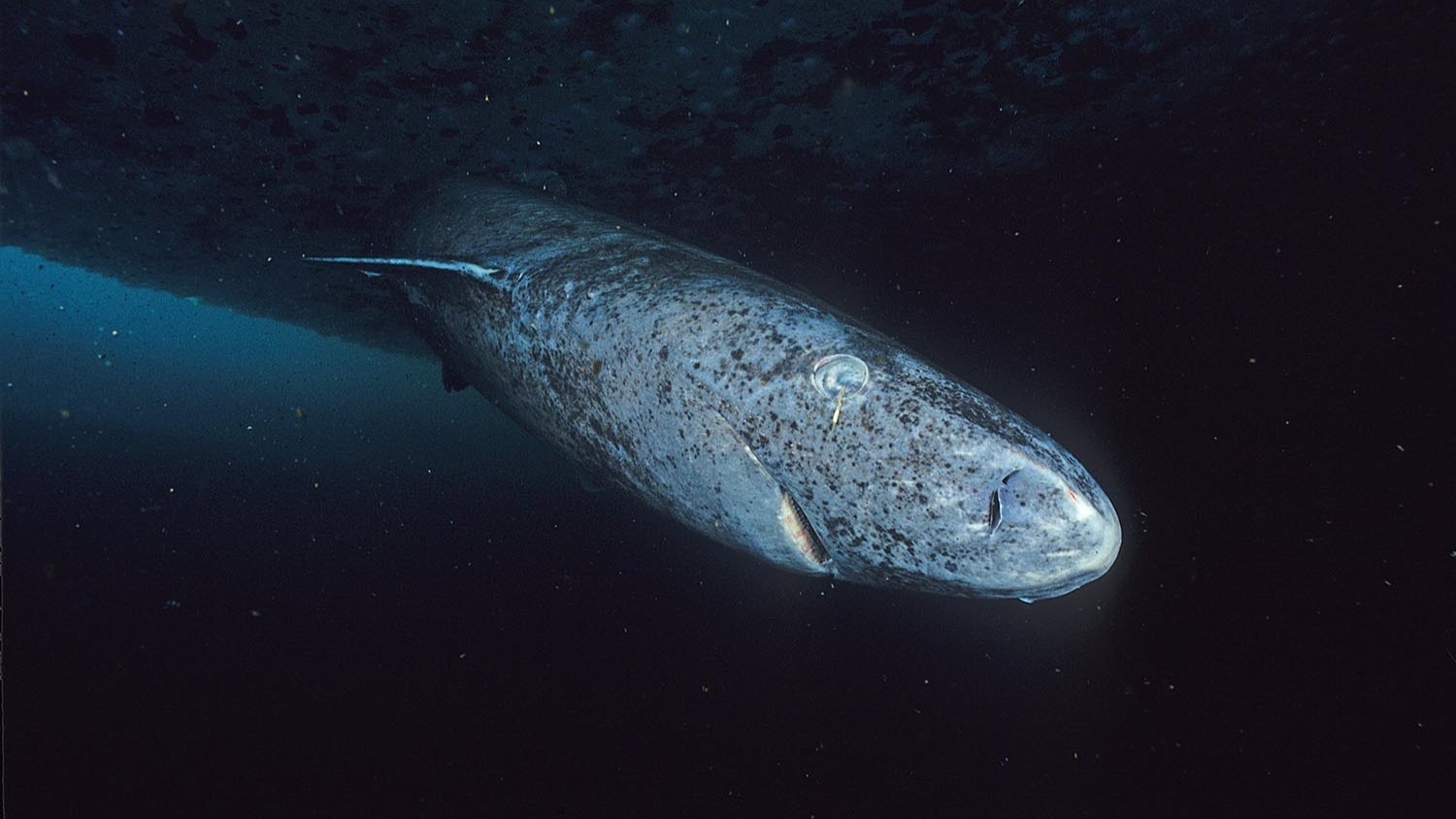
The Greenland shark widely described as 392 years old did not arrive with a birth date written into its bones. It did not have annual rings that could be counted. The number came from radiocarbon in the center of an eye lens, interpreted through a model with a very wide span of plausible ages.
The animal was a 5.02-meter female, the largest of 28 female Greenland sharks in a 2016 study published in Science. The model placed her age at 392 years, with a 95.4 percent probability range from 272 to 512 years.
That 240-year interval is not a footnote. It is the central fact needed to read the result honestly. The shark was not verified as exactly 392 years old, and the study did not prove that she reached 512.
This is one study, not settled consensus. Its measurements strongly supported extreme longevity, but the age assigned to the largest individual remained a model-based estimate.
A shark collected by accident
The research led by Julius Nielsen of the University of Copenhagen used sharks collected as bycatch during scientific fish surveys in Greenlandic waters from 2010 to 2013. They were not caught and killed specifically for an aging experiment.
All 28 were female. They ranged from 81 centimeters to 502 centimeters in total length, giving the researchers animals from very different stages of the species’ long life.
Greenland sharks are awkward animals to date. Many bony fish can be aged from growth bands in their ear stones, or otoliths. Some sharks can be studied through bands in calcified vertebrae or fin spines. Those conventional structures are poorly suited to the Greenland shark.
ScienceBlog has previously described the species’ slow growth and extraordinarily delayed maturity. The narrower question here is how much confidence the evidence allows in one especially memorable age.
The eye lens serves as a time capsule
The solution lay in the nucleus of the shark’s eye lens. Lens fibers form in layers, and the proteins at the center are produced very early in development. Unlike many tissues, that central material does not keep turning over throughout life.
Its carbon therefore retains information from around the shark’s birth. By measuring the ratio of radioactive carbon-14 in those proteins, the team could compare the result with changes in environmental radiocarbon through time.
The idea is simple to state, but marine radiocarbon is not a universal clock. Carbon can remain in deep water for long periods before entering a food web. The developing shark’s lens also receives carbon indirectly through its mother’s diet.
That “marine reservoir” effect means old ocean carbon can make a young tissue look older than it is unless the calibration accounts for where its carbon came from. The researchers used marine calibration data and information from comparable predators, but uncertainty remained.
Nuclear weapons provided the youngest time marker
Atmospheric nuclear testing during the 1950s and early 1960s sharply increased carbon-14. That global rise, known as the bomb pulse, appears in biological tissues formed after the tests and can provide a dated marker.
Only the three smallest sharks, all 220 centimeters or shorter, showed signs of the bomb pulse. This supported the conclusion that they had been born after the pulse reached the marine food web.
The marker helped confirm that body length was associated with age. It did not stamp a birth year onto the largest sharks, whose lens nuclei contained pre-bomb carbon.
For animals born centuries earlier, the radiocarbon calibration curve is relatively flat. Small differences in a measurement can therefore correspond to large differences in calendar age. The clean anchor available for the youngest sharks disappears.
How 392 became the headline number
The researchers combined the radiocarbon results with a Bayesian model of shark growth. For the largest pre-bomb animals, the model produced probability distributions rather than single certain ages.
The 5.02-meter shark’s distribution had a midpoint of 392 years. The paper reported it as 392 plus or minus 120 years, using the extent of a 95.4 percent probability range. Subtracting and adding 120 gives 272 to 512 years.
The midpoint is not necessarily a known birth year, and the two endpoints are not alternative birthdays with equal status. They mark the reported span within which the model placed most of its probability, given the data and assumptions.
This is why headlines calling the animal a “512-year-old shark” are wrong. So are historical timelines that confidently place her birth in one named year. The honest statement is that her age was estimated across a range of more than two centuries.
Why the interval is so wide
A 240-year window can sound almost useless. It is better understood as the visible consequence of several difficult problems being carried through the calculation instead of concealed.
The old portion of the radiocarbon curve changes slowly. The reservoir age of the carbon entering Arctic food webs can vary by place and depth. The relationship between length and age also has to be modeled from a sample with few very large individuals.
The two largest sharks measured 4.93 and 5.02 meters. Their midpoint estimates were about 335 and 392 years respectively, but both estimates were broad. A larger body did not provide the precision of counting rings.
A Virginia Institute of Marine Science explanation of the work made the uncertainty explicit from the beginning: 392 years was an estimate with 120 years on either side.
The lower edge still changed the record
The result remained consequential because of what happened at the bottom of the interval. Before this study, the best-known vertebrate longevity benchmark belonged to the bowhead whale, with one individual estimated at roughly 211 years.
Even 272 years, the Greenland shark’s lower bound, exceeded that by about six decades. The Smithsonian Ocean account highlighted precisely this comparison.
The study’s defensible record was therefore not “we know this shark was born 392 years ago.” It was that the evidence placed the longest Greenland shark lifespan beyond the known vertebrate record even under the cautious edge of the reported interval.
“Oldest vertebrate” also does not mean oldest animal. The ocean quahog nicknamed Ming was determined to have lived for 507 years, and some other invertebrates or colonial organisms can persist far longer. The shark record belongs to animals with backbones.
One sampled shark is not a living celebrity
The 5.02-meter animal was already dead when researchers received it as bycatch. The paper did not tag a particular shark and then announce that the same individual was still swimming centuries later.
That matters because social media posts often pair the number with an unrelated photograph and present the subject as a named ancient survivor. Most images show another Greenland shark and cannot reveal its age by appearance.
The species-level conclusion is more secure than any story attached to a viral portrait. Radiocarbon evidence from the sampled animals supports the possibility of lives measured in centuries. It does not make every large Greenland shark 400 years old.
Later work continues to examine how such animals function. A 2025 study of the Greenland shark visual system, for example, found intact retinal organization and molecular signs consistent with maintained dim-light vision. It did not independently redetermine the famous specimen’s age.
Extreme longevity makes recovery slow
The 2016 model also estimated female sexual maturity at least 156 years, with an uncertainty of 22 years on either side. That estimate was linked to a female length at maturity of roughly four meters.
This is where the result becomes more than a longevity curiosity. If females need well over a century before reproducing, losses are replaced on a timescale far longer than a fishing season, a management cycle or a human career.
The paper itself raised conservation concerns for that reason. A shark removed before maturity may represent more than a century of growth that never contributes offspring.
The maturity estimate comes from the same broad model and should not be read as a universal birthday for reproduction. Yet even substantial uncertainty leaves an exceptionally slow life history.
The uncertainty is part of the discovery
The temptation is to rescue the story by choosing one dramatic number. Say 392 years and the shark fits neatly into a historical timeline. Say 512 and the claim becomes even easier to share.
The study is more interesting when its uncertainty remains visible. Scientists found a tissue that preserves early-life carbon, used the bomb pulse to anchor the young end of the sample, and built a model for animals born before that anchor existed.
The result could not identify one exact year. It could support something broader: Greenland sharks can live for at least 272 years, and the largest animal in this sample may have lived much longer.
That is enough to establish the vertebrate longevity record without pretending the eye lens supplied a birth certificate. The 240-year window is not where the finding fails. It is where the measurement tells us how far it can honestly go.