

The paradox: In animal models there is a consistent relationship between eating less and living longer. But studies in humans find that people who are a little overweight live longest.
Last week, I introduced this paradox and offered evidence, both that lab animals live longer when they are underfed, and that humans live longer when they are overfed. In the article below, I introduce nuances and confounding factors, but in my opinion, the paradox remains unresolved.
BMI
BMI is an imperfect measure of how fat or thin someone is for his height. That’s because it is calculated with the square of height, but body volume (for a given shape) is proportional to the cube of height. The result is that tall people will have a higher BMI than shorter people with equivalent proportions of body fat. For example, BMI=20 for a person 5 feet tall means a weight of 102 pounds, an average weight for that height; whereas BMI=20 for a person 6 feet tall means a weight of 147, which is borderline emaciated.
Short people tend to live significantly longer than tall people, and the effect is substantial. Males under 5’7” live 7½ years longer than males over 6’ [ref]. This fits with the fact that short people tend to have less growth hormone in their youth. There is a genetic variant in parts of Ecuador that prevents growth hormone from transforming to IGF1 (Laron dwarfism); these people are generally about 4 feet tall and tend to live longer. From domesticated animals, we also know that small dogs live longer than large dogs, small horses longer than large horses. Between species, larger animals live longer, but within a single species, smaller animals live longer.
The height association deepens the weight paradox, because short people will tend to have a lower BMI, which we would expect to skew the association of BMI with longevity downward.
Growth Hormone and IGF1
Growth hormone (which is translated into IGF1 in the body) is genetically associated with shorter lifespan, but we have more of it when we’re young and it promotes a body type with more muscle, less fat. According to this Japanese study, IGF1 increases with weight for people who are thin, but decreases with weight for people who are fat. So maximum longevity is close to maximum IGF1.
Here are some partial explanations for the paradox.
Most variation in weight is explained by genetics, not food intake. The explanation I have proposed in the past is that the CR effect is about food intake, not genetics. And people who are congenitally stout are more likely to be restricting their calories. CR humans are not necessarily especially thin.
The CR effect is proportionately smaller in long-lived humans than in short-lived rodents or shorter-lived worms and flies. [ref] If life extension via CR evolved to help an animal survive a famine, then it seems reasonable that the benefit should be limited to a few years, because that is as long as most famines in nature are likely to last.
The CR effect may be due to intermittent fasting rather than total calorie intake. Traditional CR experiments conflate intermittent fasting with overall calorie reduction, because food is provided in a single daily feeding, and hungry rodents gobble it up, then go hungry for almost 24 hours. More recent experiments attempt to separate the effect of limited-time eating from the effect of calorie reduction, and the general conclusion is that both benefit longevity. It may be that humans who are skinny tend to graze all day, while people with a comfortable amount of fat more easily go for hours at a time without eating.
Mice carry less fat, have less food craving, and have better gut microbiota if they are fed at night rather than during the day [ref]. Mice are active nocturnally; so translating to humans, it probably means that we should eat in the morning. Conventional wisdom is that eating earlier in the day is better for weight loss and health [ref], but I know of no human data on mortality or life span. This classic study in mice [1986] found caloric restriction itself was the only thing affecting lifespan, and there was no difference whether the mice were fed night or day, in three feedings or one.
Smokers tend to be thinner than non-smokers, but they don’t live longer for reasons that have to do with smoking, not weight. So this is a partial explanation why heavier BMI might be associated with longer lifespan. But note that the recent Zheng’s Ohio State study claimed there was no change in the best weight for longevity when correction was introduced for smoking.
Cachexia is a “wasting” disorder that causes extreme weight loss and muscle atrophy, and can include loss of body fat. This syndrome affects people who are in the late stages of serious diseases like cancer, HIV or AIDS, COPD, kidney disease, and congestive heart failure (CHF). [healthline.com] If cachexia subjects are not removed from a sample, it can strongly bias against weight loss, because once cachexia sets in, life expectancy is very short. But the Zheng study was based on Framingham data, collected annually over the latter half of a lifetime; Cachexia is not expected to be a significant factor.
Timing artifact – The Framingham study covers a 74-year period in which BMI is increasing and also lifespan is increasing, probably for different reasons. The younger Framingham cohort is living ~4 years longer than the older cohort and is ½ BMI point heavier. This could create an illusion that higher BMI is causing greater longevity. However, the Ohio State study made some effort to pull this factor out. Greater lifespan is associated with gradually increasing BMI, and this is true separately in both cohorts.
Differential effects on CVD and Cancer This chart (from Zheng) shows how the mortality burden of cardiovascular disease has decreased over the last century, but not so cancer.
But CV disease risk increases consistently with BMI, while cancer risk, not so much (also from Zheng):

| These numbers in parentheses are odds ratios from a Cox proportional hazard model. What they means is that a person in the Lower-Normal weight group had 20% less chance of getting heart disease compared to someone of the same age in the Normal-Upward group, but a 60% increased chance of getting cancer. These appear to be large, concerning numbers. But remember that the underlying probabilities are all increasing exponentially with age. Translated into years of lost life, 60% greater probability of cancer is only 1 year of life expectancy at age 50. (60% greater overall mortality would subtract 4½ years from life expectancy.) In my experience, hazard ratios in the range 0.7 to 1.5 don’t necessarily mean anything, because of the difficulties in interpreting data. The numbers in parenthesis after 1.60 in the above table (1.12 — 2.30) mean that statistical uncertainty alone is a range from 1.12 to 2.30.There are plenty of large effects with hazard ratios of 3 or more. For comparison, the hazard ratio for pack-a-day smokers getting lung cancer is 27. |
Zheng’s study found a longevity disadvantage to being underweight, and it was exclusively due to a higher cancer risk. In fact, incidence of cardiovascular disease among the lowest BMI class was lowest (0.8); but their cancer risk more than made up for it (1.6).
This means that as time goes on and most Americans are getting heavier, their risk of dying from CVD is blunted by improved technology. The mortality risk from CVD is down by 40% in this century [NEJM], while the cancer risk is unchanged [CDC]. So people are dying of cancer who would have died of CVD in previous generations.
This means that low BMI has less benefit for longevity than it used to have, and the trend over time tends to exaggerate the appearance that higher weight is protective against all-cause mortality.
Is it true that cancer risk does not go up with BMI?
The Framingham result is puzzling and difficult to reconcile with a well-established relationship between higher BMI and higher cancer risk. This review by Wolin [2010] finds a modest increase in risks of all common types of cancer associated with each 5-point gain in BMI. (The RR numbers are comparable to hazard ratios above.)

Lung cancer is the big exception, and Wolin explains the inverse relationship with BMI by the fact that people smoke to avoid gaining weight. This would suggest a resolution to the conflict with Zheng’s study, but for the fact that Zheng explicitly corrects for smoking status and finds it makes no difference at all — a result which is puzzling in itself.
Alzheimer’s Disease is the third leading cause of death, and the corresponding story is more complicated. Lower weight in middle age seems to be mildly protective, while it is certainly not protective in the older years when AD is most prevalent.
“Hazard ratios per 5-kg/m2 increase in BMI for dementia were 0.71 (95% confidence interval = 0.66–0.77), 0.94 (0.89–0.99), and 1.16 (1.05–1.27) when BMI was assessed 10 years, 10-20 years, and >20 years before dementia diagnosis.” [ref]
This, too, is unexpected in light of previous consensus. Alzheimer’s Dementia has been recast as Type 3 Diabetes, because of its strong association with insulin metabolism. Overweight is supposed to be the greatest life-style risk factor for diabetes. When this study [2009] out of U of Washington found that high BMI is protective against dementia, the authors were unwilling to draw the standard causal inference, so they conjectured instead that weight loss is a consequence of AD’s early stage.
There may be a better explanation hidden in their data. AD is the most common cause of dementia, but vascular dementia, a separate etiology, accounts for roughly ⅓ of cases in the Kame data set: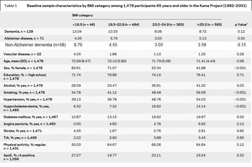
There is a suggestion here that higher BMI protects against vascular dementia, but not against AD.
From you, my readers
Here are some of the suggestions offered in the comment section of last week’s blog:
- Fat people are happier. I don’t doubt that happiness has a lot to do with longevity but a lot of overweight is due to compulsive eating by people who are not happy with their lives. Obesity is associated with lower socio-economic status, and lower SES is independently associated with shorter lifespan and lower life satisfaction.
- Higher BMI can mean more muscle mass, not necessarily more fat mass. Good point. I don’t know how big a factor this is.
- This study [BMJ 2016] found greatest longevity for BMI in the range 20-22. I take your point that the larger studies with longer follow-up tend to report lower optimal BMI. The BMJ study is a meta-analysis of a huge database covering 9 million subjects.
- Dean Pomerleau writes at the CR Society web page about brown fat, cold resistance, and greater longevity.
- Thin people have greater insulin sensitivity, which can lead to glucose going into cells instead of being stored as fat. This is interesting, and deserves more follow-up. But good insulin sensitivity also means lower blood sugar, so it’s not obvious to me which direction the effect ought to go.
- I was grateful for a pointer to Valter Longo’s recent work, recommending that time-restricted eating becomes counterproductive after about 13 hours a day of fasting. Longer fasts several times a year are still highly recommended.
- Paul Rivas is my go-to authority on weight, and he recommended this 2015 study, which emphasizes the paradox as I describe it.
- This study out of Emory U [2019] recommends different diets for different BMI groups for minimizing inflammation.
What story does methylation tell?
Aside from mortality statistics, I regard methylation age as the most reliable leading indicator we have. I’ll end by reviewing data on BMI and methylation age.
The Regicor Study [2017] looked for methylation sites associated with obesity. They reported 97 associated with high BMI and an additional 49 associated with large waistline. I compared their lists with my list of methylation sites that change most consistently with age. There was no overlap. What I learn from this is that there is no association with genetically-determined weight and longevity. If you were born with genes that make you gain weight, there is a social cost to be paid in our culture, but there is no longevity penalty.
Horvath [2014] did not discern a signal for obesity with the original 2013 DNAmAge clock, except in the liver where the signal was weak, amounting to just 3 years for the difference between morbidly obese and normal weight. But a few years later with 3 different test groups [2017], a moderate signal was found, as expected, linking higher BMI to greater DNAmAge acceleration. (Age acceleration is just the difference between biological age as measured by the methylation clock and chronological age by the calendar.)
This study [2019] from the European Lifespan Consortium found a modest increased mortality from obesity, corresponding to less than a year of lost life by most measures, based on two Horvath clocks and the Hannum clock. This Finnish study [2017] found a small association between higher BMI and faster aging in middle-aged adults, but not in old or young adults.
This study from Linda Partridge’s group [2017] found a strong benefit of caloric restriction on epigenetic aging—in mice, not in humans.
 The bottom line
I’ve had a good time with this project, seeking explanations for the paradox, and I’ve passed along some interesting associations, but in the end, the essential paradox remains. I don’t know why the robust association of caloric restriction with longevity doesn’t lead to a clear longevity advantage in humans for a lower BMI. My strongest insight is that the largest determinants of BMI are genetic, not behavioral, and the genetic contribution to weight has no effect on longevity. But what do I make of the fact that life expectancy in the US has risen by a decade over my lifetime [ref] even as BMI has increased 5 points.
Discussion
160 reader comments
Imported threads are marked Archive. New comments are welcome and moderated for spam.
In stem cell therapy adult stem cells often get harvested from fat tissue. I don't know why fat seems to have a higher stem cell concentration but it could perhaps help explaining the BMI paradox. The net effect of (a) the risks associated with a higher BMI and (b) the renegerative benefits of the stem cell population is positive.
Josh,
at your bottom line you said:
" I don’t know why the robust association of caloric restriction with longevity doesn’t lead to a clear longevity advantage in humans for a lower BMI"
I do not say what follows is correct but, what about the De Gray published claim aroun 2007 that selection for the CR adaptation to famine depends on the relative length of famines in the wild (rarely longer than one year or two) vs. the life span of the species considered, which would explain why CR clearly (no reasonable doubt after 90 years of experimentation specially from the 1970's mainly at Texas medical? center) increases longevity in (short-lived species) rodents but not in (long-lived species) humans?
The solution to this problem CANNOT BE studies in humans. You will lose your peecious time and money studying humans with their too many confounding factors impossible to statistically eliminate (those who claim they do are lying fakes only due to own corporative interests, medical drs., big pharma in health issues, same for fake siciologists or economists etc who say their disciplines are "scientific"! But they are not! There are only 4 Sciences:
2 BEST HARD ONES: PHYSICS AND CHEMISTRY
and Two Less Hard and derived from the 2 first ones: GEOLOGY and BIOLOGY
Coming back to CR, then, how to solve Josh's dilemma?
In my opion the best is to go to intermediate longevity species....and that huge effort has already been done and WILL NEVER BE REPEATED ( concernibg rhesus this last sentence is rge only thing in which I agree with the clearly biased for his e onomic interests in 2013 Aubrey declared at Madrid Phylosophy Faculty UCM invited by me extending his presentation to 2,000 UCM Proffessors!
CR in rhesus macaques (max. longevity around 35 years), 2 experiments published years ago
1. Aubrey (scandalously biased) only considered Mattison et al. Nature 2012 (GOVERNMENTAL CONTROLLED NIH STUDY).
2. Richard Weindruch Wisconsin University Primate Center directed experiment (I consider Richard the highest scientific "authority" worldwide withou any discussion posible): Coleman et al. Science (2009)
Both studies agree that CR in Rhesus significantly decrease/delay incidence of degenerative diseases. But only Richard experiment showed inprovement in longevity (experiment not finished to max. In Coleman Science 2009.).
I suggest you to read both papers plus a third one (in 2014) signed by both Coleman and Mattisson trying to ascertain why longevity outcome was different in both studies.
After your detailed reading FROM TOP TO BOTTOM. Please read both papers IN FULL , NOT ONLY THE ABSTRACT! (you will not be so tired it is only 2 papers: I read 10,000 IN FULL in my whole life and perhsps that is why finally I discovered the gene ndufv2 responsible for mitROSp and around 1/3? Zof aging rate?
I did read Mattison and Coleman papers in full and more than once in my life. In my opion both experiments have design problems but....if I must choose one.....I CHOSE COLEMAN .
The implication is that in my opion the Aubrey de Grey argument about CR impossibly selecting for increased human longevity is flawed.
No doubt De Grey is intelligent enough to understand that he must dismiss/ignore Coleman' s paper (as he scandalously did at Madrid even after me -co Chair and co-organizer of the of his talk recorded in TV still now available on the web) . If he considers Coleman, then his ONLY THEORETICALLY BASED prejudice falls down.
And do not forget that Aubrey de Grey IS NOT A SCIENTIST he is, as he always claimed while (unfairly) despising both geriatricians and GERONTOLOGISTS! , AN ENGINEER!
Of course he is that! And that is why he is so wrong about Aging, an obiously biological and thus scientific subjetc
Engeneers please better stay doing radios, bridges and the like but not give opiion on animals which they absolutely ignore how ate they builded up inside. (Aubrey is an ignorant without even a biology degree. Do not give money to that cheater. That is my uninterested advice.
@Gustavo Barja
sorry for replying so late to your comment on appreciating my hypothesis on aging. I am no scientist, but a layman following the anti aging field for the last 10 years.
Here is my take again.
Multi cellular organisms are governed by gene networks right from embryogenesis till death. I say death because i feel aging is also controlled by a gene network, which comes in to being at puberty. I also feel that morphogenesis does not end at birth, it continues till the end of the pubertal phase. Morphogenetic pace not phase varies across species with varying intervals of growth pace.
A gene network has function not individual genes, an individual gene can be part of many gene networks in the same organisms(for e.g serotonin). it is the gene network that morphs at various tissue growth milestones or organ boundary milestone and acquires different forms and function at those milestones. It does this by inhibiting some of its member genes and/or adding new genes to its network.
Aging is one such network which is activated at the morphogenetic milestone of puberty. The activation of this network, results in a program which controls
1 :the repair functions of the cell at the individual level, such as autophagy,dna repair, protein digestion, mitochondria repair etc.
2: the recruitment of the immune system in response to cell damage and
3: the renewal provided by resident stem cells.
The gene network which was in control of the above functions during the growth and the final morphogenetic turn at puberty, is modified by activation of some unidentified genes, which in turn inhibit some genes, which played a key role in the previous iteration of the network. Due to this, the whole network modifies itself to implement the aging program.
And therefore, various genes which were found to be beneficial in the previous avatar of the network turn deleterious in the new network, leading to the so called phenomenon 'antagonistic pleiotropy'.
Various supplements which have proven to be beneficial are just perturbing small parts of the overall network. However, with the epigenetic clock showing lock step epigenetic modification across various organs and tissues in the organism. the presence of signalling factors in the communication medium can be hypothesized. Hence plasma exchange/dilution are probably effective because the perturb the whole network.
Also according to me partial reprogramming is an unnatural way of changing the dynamics of the aging gene network by introducing elite transcription factors, which supersede the key transcription factors controlling the aging network.
The recent paper on albumin
Young and Undamaged rMSA Improves the Longevity of Mice
'125 mg/mL of rMSA dissolved in saline was i.v. injected slowly. Mice were weighed before each injection to calculate the dosage, with saline served as the negative control. Mice were injected with 1.5 mg rMSA per gram of mouse body weight and isometric saline every 3 weeks as indicated.'
Actuals according to various papers
average body weight of mice 30 g
average plasma volume of mice 1.6 ml
average concentration of Albumin in mice 25 mg/ml
average total albumin in mice 40 mg
The paper also does not mention clearly the total volume of saline with albumin injected per mouse. It mentions 125 mg/ml was injected, whereas average concentration of Albumin in mice 25 mg/ml. But in the same paragraph it mentions 'Mice were injected with 1.5 mg rMSA per gram of mouse body weight', which comes to average 45 mg albumin injected.
Whereas 125 mg/ml concentration comes to 187.5 mg total concentration in plasma of 1.5 ml.
The latest and greatest anti-aging intervention?
Glycine and N-acetylcysteine (GlyNAC) supplementation in
older adults improves glutathione deficiency, oxidative
stress, mitochondrial dysfunction, inflammation, insulin
resistance, endothelial dysfunction, genotoxicity, muscle
strength, and cognition: Results of a pilot clinical trial
https://www.ncbi.nlm.nih.gov/pmc/articles/PMC8002905/pdf/CTM2-11-e372.pdf
"GlyNAC supplementation for 24 weeks in OA corrected RBC-GSH deficiency, OxS, and mitochondrial dysfunction; and improved inflammation, endothelial dysfunction, insulin-resistance, genomic-damage, cognition, strength, gait-speed, and exercise capacity; and lowered body-fat and waist circumference. However, benefits declined after stopping GlyNAC supplementation for 12 weeks. Supplementing GlyNAC in aging humans could be a simple and viable method to promote health and warrants additional investigation"
Thought the oxidation theory or aging was dead? Non essential amino acids having profound anti-aging benefits?
For a 70kg person, 7g glycine/day and 9.3 g NAC/day.
Well the strict oxidation hypothesis is clearly wrong as that would imply higher metabolism would inevitably result in lower lifespan. But there are known exceptions to this rule. What is now known I think goes by the name membrane pacemaker modification, or something like that, it shows that the peroxidation index of membranes is modified in species with longer than expected lifespan for their metabolism. Species make membranes more oxidation resistant if they are longer lived for a particularly high metabolism.
I think this is also the case in insects, when comparing long lived queens vs short lived workers. Queens membranes are more peroxidation resistant than worker membranes. Keep in mind that some insect colonies are rumored to have up to 100x difference in lifespan between queens and workers, despite having identical genomes. Showing how changes to regulation of gene expression can lead to vast differences in lifespan.
What I'd need to research is how it works for neurons, given I've heard they tend to have a lot of polyunsaturated lipids in their membranes, which would presumably be easily oxidable and a very bad idea given their extremely high metabolism. Yet neurons are said by some to be biologically immortal in the right environment. They live as long as the animal lives without dividing at extreme metabolic rates, how they accomplish this is a good question.
How a body can have immortal nondividing cells at high metabolism yet the rest of the dividing mostly lower metabolism body ages, aka less oxidation and damage due to lower metabolism, is a good question.
Darian it is you who is wrong whe you ignorantly critizize MFRTA, because you are assuming that FRL% is a constant which is not, absolutely not!!!!!. I wrote a whole paper (Barja Rejuv. Research 2007) to explain this to those who do not understand how the mitochondrial electron transport chain works, the 100% of scientists at USA since Britton Chance (PA) died. It is clear that you are one more if those ignorants. It is terrible how much ignorance pervades USA science due to you being dominated by FASHION!. SCIENCE IS NOT FASHION. AND BRITTON WAS A GOD. YOU LODT HIM AND YOU WERE LEFT IN ABSOLUTE IGNORANCE ON MITICHONDRIAL FUNCTION. I SPEAK OF FUNCTIONAL WELL COUPLED (ALIVE!!!) RESPIRING MITOCHONDRIA. CHRISTOPH RICHTER A GERMAN TAUGHT ME HOW TO OBTAIN THEM FROM FRESH TISSUE IN 1994. AT THAT TIME NO ONE IN THE WHOLE USA KNEW HOW TO BREAK THE CELL AND TAKE OUT OF IT WELL COUPLED FUNCTIONAL MITOCHONDRIA. NOW YOU HAVE AT BUCK INSTITUTE ANOTHER EUROPEAN (OF COURSE!!!) WHO KNEW HOW TO DO THAT, THE KING OF H+ LEAK MARTIN BRAND FORMERLY PROFFESSOR OF BIOCHEMISTRY AT CAMBRIDGE UNIVERSITY UK. YOU BOUGHT HIM WITH YOUR VALUE: DOLLARS (HE RENOUNCED TO HIS CAMBRIDGE PROFESSIRSHIP!). HE WILL RETURN TO ENGLAND WITH HIS MILLIONAIRE RETAIRMENT. THAT IS WHAT HE WANTED FROM YOU IN INTERCHANGE FOR WHAT HE GAVE YOU CONCERNING FUNCTIONAL MITOS. I HELPED MARTIN TO LEARN HOW TO PROPERLY MEASURE MITROSp (I SENT MY STUDENT SUSANA CADENAS WHO DID HER PhD with ME AT MADRID TO DO A 4 YEARS LONG PISTDOC WITH MARTIN AT CAMBRIDGE UK....
What are you talking about?
Mitochondria free radical has an effect. But it has clearly been seen that altering membrane composition to reduce oxidation is enough to essentially do away with the issue. Humans are said to have even more oxidation resistant mitochondrial membranes, iirc.
Now some species essentially show little to no signs of aging, and then drop dead, probably due to programmed death program. Others even have unlimited lifespans as far as we can tell. Is there a movement of additional mitochondrial genes to the nucleus, not as far as I know.
Our rodent ancestor likely had a lifespan of a few years, or primate ancestor a few decades at most, yet we live over 115 years. Few genetic mutations allowed this.
With regards to the brain with 115+ year long lived cells, that can probably last for centuries while keeping high metabolism, maybe their mitochondrial membranes are even more oxidation resistant than other tissues. Their external membranes are said to have lots of polyunsaturated lipids, iirc. Maybe they have protections for that. But perhaps they deal with the issue by dealing with the mitochondria membrane.
Know this mice neurons last OVER TWICE AS LONG AS MICE THEMSELVES DO when transplanted into another host, and researchers have speculated these cells to be biologically immortal.
9.3 grams of NAC per day seems like an awful lot, especially when we keep in mind that NAC has pro-oxidant effects at large doses. It's certainly an interesting study and the results cannot be ignored.
...in addition, we need to see the same study conducted on HEALTHY AND FIT OAs without insulin resistance.
I'm wondering, if changing diet, incorporating exercise and fasting would have led to similar health benefits, considering that the room for improvement is so vast for these individuals.
No it doesn't - the inflammatory cytokines caused by elevated ROS mean you can't gain muscle without first lowering ROS (if you are in that aged state). But it is a fine balance, too low ROS and autophagy doesn't work.
It is a mystery why to me why this paper has got so much attention. Wulf Droge (RIP) was talking about this in the early 2000s.
Check out my two new updates for my aging blogpost>>>
link>> https://jefftbowles.com/aging-is-programmed/
title >> NEW STUDY DISPROVES ALL MAINSTREAM THEORIES OF AGING-AND REVEALS THE NEW: PROGRAMMED LOSS OF CELLULAR DIFFERENTIATION THEORY OF AGING
Updtes>>> the coolest one first>>>
Update #13
I wrote in my 1998 paper > ” Cancer, in a broad sense, may simply be a cell returning to its earlier, primitive, immortalized, state. It should not be very surprising that a mortal life form that evolved from a previously immortalized life form could spontaneously become immortalized through loss of some type of control. However, if the mortal life form had evolved from mortal ancestors, spontaneous immortalization would seem to be quite a miracle indeed.” see comment in full context at the end of this update .
Well, well, well>> I have been reading all the abstracts in Pub Med that contain the term “Yamanaka Factors” and what have I found? The cancer cells are basically just malfunctioning de-differentiated embryonic stem cells. And yes it seems very likely they are simply a reemergence of our oldest ancestors…Single cell life that lived before the age of oxygen. Both cancer cells and embryonic stem cells can dviide indefinitely (immortal). Both of the them do not use oxygen for energy even when oxygen is present but rather switch to an anaerobic form of glycolysis for energy ! What follows are the interesting abstracts that show how this view of cancer being a reversion of cells to their most primitve state seems to be correct:
The role of pluripotency factors to drive stemness in gastrointestinal cancer
Abstract
A better molecular understanding of gastrointestinal cancers arising either from the stomach, the pancreas, the intestine, or the liver has led to the identification of a variety of potential new molecular therapeutic targets. However, in most cases surgery remains the only curative option. The intratumoral cellular heterogeneity of cancer stem cells, bulk tumor cells, and stromal cells further limits straightforward targeting approaches. Accumulating evidence reveals an intimate link between embryonic development, stem cells, and cancer formation. In line, a growing number of oncofetal proteins are found to play common roles within these processes. Cancer stem cells share features with true stem cells by having the capacity to self-renew in a de-differentiated state, to generate heterogeneous types of differentiated progeny, and to give rise to the bulk tumor. Further, various studies identified genes in cancer stem cells, which were previously shown to regulate the pluripotency circuitry, particularly the so-called “Yamanaka-Factors” (OCT4, KLF4, SOX2, and c-MYC). However, the true stemness potential of cancer stem cells and the role and expression pattern of such pluripotency genes in various tumor cell types remain to be explored. Here, we summarize recent findings and discuss the potential mechanisms involved, and link them to clinical significance with a particular focus on gastrointestinal cancers.
The oncogene c-Jun impedes somatic cell reprogramming
Jing Liu 1, Qingkai Han 1, , Duanqing Pei 2
Oncogenic transcription factors are known to mediate the conversion of somatic cells to tumour or induced pluripotent stem cells (iPSCs).
EMBO Rep
. 2014 Mar;15(3):244-53.
Dedifferentiation and reprogramming: origins of cancer stem cells
Abstract
Regenerative medicine aims to replace the lost or damaged cells in the human body through a new source of healthy transplanted cells or by endogenous repair. Although human embryonic stem cells were first thought to be the ideal source for cell therapy and tissue repair in humans, the discovery by Yamanaka and colleagues revolutionized the field. Almost any differentiated cell can be sent back in time to a pluripotency state by expressing the appropriate transcription factors. The process of somatic reprogramming using Yamanaka factors, many of which are oncogenes, offers a glimpse into how cancer stem cells may originate. In this review we discuss the similarities between tumor dedifferentiation and somatic cell reprogramming and how this may pose a risk to the application of this new technology in regenerative medicine.
J Cell Sci
. 2013 Aug 15;126(Pt 16):3638-48.
The reprogrammed pancreatic progenitor-like intermediate state of hepatic cells is more susceptible to pancreatic beta cell differentiation
Abstract
Induced pluripotent stem cells (iPSCs) hold great promise for cell therapy. However, their low efficiency of lineage-specific differentiation and tumorigenesis severely hinder clinical translation. We hypothesized that reprogramming of somatic cells into lineage-specific progenitor cells might allow for large-scale expansion, avoiding the tumorigenesis inherent with iPSCs
Expert Rev Anticancer Ther
. 2021 Apr 8.
Pluripotency inducing Yamanaka factors: role in stemness and chemoresistance of liver cancer
Abstract
Introduction: Liver cancer is a major cause of mortality and is characterized by the transformation of cells into an uncontrolled mass of tumor cells with many genetic and epigenetic changes, which lead to the development of tumors. A small subpopulation of cell population known as Cancer Stem Cells (CSCs) is responsible for cancer stemness and chemoresistance. Yamanaka factors [octamer-binding transcription factor 4 (OCT4), SRY (sex-determining region Y)-box 2 (SOX2), kruppel like factor 4 (KLF4), and Myelocytomatosis (MYC); OSKM] are responsible for cancer cell stemness, chemoresistance, and recurrence.
Biochem Biophys Res Commun
. 2019 Sep 17;517(2):324-329.
Silencing of the transcription factors Oct4, Sox2, Klf4, c-Myc or Nanog has different effect on teratoma growth
Abstract
Induced pluripotent stem cells (iPSC) have a great potential, but their clinical application depends on finding strategies to abolish their tumorigenic potential. The use of Oct4, Sox2, Klf4, c-Myc and Nanog to generate iPSC demonstrated the already known importance of these genes to maintain stemness. Therefore, the presence of these genes is responsible for iPSC-derived teratomas. Similar to iPSC, P19 teratocarcinoma cell line also has characteristics of embryonic carcinoma cells and the ability to differentiate into many cell types. We separately silenced the transcription factors Oct4, Sox2, Klf4, c-Myc and Nanog in P19 cells and measured the impact of this silencing in vivo. All silenced cells generated tumors when injected in immunosuppressed mice, but silencing of Oct4, Sox2 and Klf4 generated mainly teratomas with mesoderm tissue. Our results suggest that downregulation of these transcription factors is not enough to avoid the formation of teratomas, but their silencing affect their differentiation potential.
Oncogene
. 2019 Aug;38(34):6226-6239.
Epigenetic reprogramming of primary pancreatic cancer cells counteracts their in vivo tumourigenicity
Abstract
Pancreatic ductal adenocarcinoma (PDAC) arises through accumulation of multiple genetic alterations. However, cancer cells also acquire and depend on cancer-specific epigenetic changes. To conclusively demonstrate the crucial relevance of the epigenetic programme for the tumourigenicity of the cancer cells, we used cellular reprogramming technology to reverse these epigenetic changes. We reprogrammed human PDAC cultures using three different techniques – (1) lentivirally via induction of Yamanaka Factors (OSKM), (2) the pluripotency-associated gene OCT4 and the microRNA mir-302, or (3) using episomal vectors as a safer alternative without genomic integration. We found that induction with episomal vectors was the most efficient method to reprogram primary human PDAC cultures as well as primary human fibroblasts that served as positive controls. Successful reprogramming was evidenced by immunostaining, alkaline phosphatase staining, and real-time PCR. Intriguingly, reprogramming of primary human PDAC cultures drastically reduced their in vivo tumourigenicity, which appeared to be driven by the cells’ enhanced differentiation and loss of stemness upon transplantation. Our study demonstrates that reprogrammed primary PDAC cultures are functionally distinct from parental PDAC cells resulting in drastically reduced tumourigenicity in vitro and in vivo. Thus, epigenetic alterations account at least in part for the tumourigenicity and aggressiveness of pancreatic cancer, supporting the notion that epigenetic modulators could be a suitable approach to improve the dismal outcome of patients with pancreatic cancer.
Methods Mol Biol
. 2019;1916:249-261.
Reprogramming of Human Melanocytes and Melanoma Cells with Yamanaka Factors
Abstract
The expression of Yamanaka factors (Oct3/4, Klf-4, Sox-2, c-Myc) can reprogram cancer cells to a pluripotent stage. This may cause the removal of their epigenetic memory and result in altered tumorigenicity. Various studies in the literature have shown that cancer cell reprogramming is a potential tool to study disease progression or discover novel therapeutic or diagnostic markers in cancer research. In this chapter, we aim to introduce the cancer cell reprogramming protocol in detail by using human melanocytes and melanoma cell lines, and Sendai viral vectors encoding Yamanaka factors have been used to reprogram cells. Representative results are discussed and important notes have been summarized in order to point out important steps during cancer cell reprogramming.
J Biomed Sci
. 2018 Jul 19;25(1):57.
Incomplete cellular reprogramming of colorectal cancer cells elicits an epithelial/mesenchymal hybrid phenotype
Abstract
Background: Induced pluripotency in cancer cells by ectopic expression of pluripotency-regulating factors may be used for disease modeling of cancers. MicroRNAs (miRNAs) are negative regulators of gene expression that play important role in reprogramming somatic cells. However, studies on the miRNA expression profile and the expression patterns of the mesenchymal-epithelial transition (MET)/epithelial-mesenchymal transition (EMT) genes in induced pluripotent cancer (iPC) cells are lacking.
Methods: iPC clones were generated from two colorectal cancer (CRC) cell lines by retroviral transduction of the Yamanaka factors. The iPC clones obtained were characterized by morphology, expression of pluripotency markers and the ability to undergo in vitro tri-lineage differentiation. Genome-wide miRNA profiles of the iPC cells were obtained by microarray analysis and bioinformatics interrogation. Gene expression was done by real-time RT-PCR and immuno-staining; MET/EMT protein levels were determined by western blot analysis.
Results: The CRC-iPC cells showed embryonic stem cell-like features and tri-lineage differentiation abilities. The spontaneously-differentiated post-iPC cells obtained were highly similar to the parental CRC cells. However, down-regulated pluripotency gene expression and failure to form teratoma indicated that the CRC-iPC cells had only attained partial pluripotency. The CRC-iPC cells shared similarities in the genome-wide miRNA expression profiles of both cancer and pluripotent embryonic stem cells. One hundred and two differentially-expressed miRNAs were identified in the CRC-iPC cells, which were predicted by bioinformatics analysis be closely involved in regulating cellular pluripotency and the expression of the MET/EMT genes, possibly via the phosphatidylinositol-3 kinases-protein kinase B (PI3K-Akt) and transforming growth factor beta (TGF-β) signaling pathways. Irregular and inconsistent expression patterns of the EMT vimentin and Snai1 and MET E-cadherin and occludin proteins were observed in the four CRC-iPC clones analyzed, which suggested an epithelial/mesenchymal hybrid phenotype in the partially reprogrammed CRC cells. MET/EMT gene expression was also generally reversed on re-differentiation, also suggesting epigenetic regulation.
Conclusions: Our data support the elite model for cancer cell-reprogramming in which only a selected subset of cancer may be fully reprogrammed; partial cancer cell reprogramming may also elicit an epithelial-mesenchymal mixed phenotype, and highlight opportunities and challenges in cancer cell-reprogramming.
Biochim Biophys Acta Rev Cancer
. 2018 Jan;1869(1):1-10.
Deubiquitylating enzymes as cancer stem cell therapeutics
Abstract
The focus of basic and applied research on core stem cell transcription factors has paved the way to initial delineation of their characteristics, their regulatory mechanisms, and the applicability of their regulatory proteins for protein-induced pluripotent stem cells (protein-IPSC) generation and in further clinical settings. Striking parallels have been observed between cancer stem cells (CSCs) and stem cells. For the maintenance of stem cells and CSC pluripotency and differentiation, post translational modifications (i.e., ubiquitylation and deubiquitylation) are tightly regulated, as these modifications result in a variety of stem cell fates. The identification of deubiquitylating enzymes (DUBs) involved in the regulation of core stem cell transcription factors and CSC-related proteins might contribute to providing novel insights into the implications of DUB regulatory mechanisms for governing cellular reprogramming and carcinogenesis. Moreover, we propose the novel possibility of applying DUBs coupled with core transcription factors to improve protein-iPSC generation efficiency. Additionally, this review article further illustrates the potential of applying DUB inhibitors as a novel therapeutic intervention for targeting CSCs. Thus, defining DUBs as core pharmacological targets implies that future endeavors to develop their inhibitors may revolutionize our ability to regulate stem cell maintenance and differentiation, somatic cell reprogramming, and cancer stem cells.
Biochim Biophys Acta Mol Cell Res
. 2017 Jul;1864(7):1359-1369.
Transdifferentiation and reprogramming: Overview of the processes, their similarities and differences
Abstract
Reprogramming, or generation of induced pluripotent stem (iPS) cells (functionally similar to embryonic stem cells or ES cells) by the use of transcription factors (typically: Oct3/4, Sox2, c-Myc, Klf4) called “Yamanaka factors” (OSKM), has revolutionized regenerative medicine. However, factors used to induce stemness are also overexpressed in cancer. Both, ES cells and iPS cells cause teratoma formation when injected to tissues. This raises a safety concern for therapies based on iPS derivates. Transdifferentiation (lineage reprogramming, or -conversion), is a process in which one mature, specialized cell type changes into another without entering a pluripotent state. This process involves an ectopic expression of transcription factors and/or other stimuli. Unlike in the case of reprogramming, tissues obtained by this method do not carry the risk of subsequent teratomagenesis.
Iran J Basic Med Sci
. 2016 Oct;19(10):1131-1135.
Linc-ROR and its spliced variants 2 and 4 are significantly up-regulated in esophageal squamous cell carcinoma
Abstract
Objectives: Similar characteristics of molecular pathways between cellular reprogramming events and tumorigenesis have been accentuated in recent years. Reprogramming-related transcription factors, also known as Yamanaka factors (OCT4, SOX2, KLF4, and c-MYC), are also well-known oncogenes promoting cancer initiation, progression, and cellular transformation into cancer stem cells. Long non-coding RNAs (lncRNAs) are a major class of RNA molecules with emerging roles in stem cell pluripotency, cellular reprogramming, cellular transformation, and tumorigenesis. The long intergenic non-coding RNA ROR (lincRNA-ROR, linc-ROR) acts as a regulator of cellular reprograming through sponging miR-145 that normally negatively regulates the expression of the stemness factors NANOG, OCT4, and SOX2.
Stem Cells
. 2016 Nov;34(11):2613-2624.
Positive Feedback Loop of OCT4 and c-JUN Expedites Cancer Stemness in Liver Cancer
Abstract
The network of stemness genes and oncogenes in human patient-specific reprogrammed cancer stem cells (CSCs) remains elusive, especially in liver cancer. HepG2-derived induced pluripotent stem cell-like cells (HepG2-iPS-like cells) were generated by introducing Yamanaka factors and the knockdown vector shTP53. They exhibited features of stemness and a higher tumorigenesis after xenograft transplantation compared with HepG2 cells. The cancerous mass of severe combined immunodeficiency (SCID) mice derived from one colony was dissected and cultured to establish reprogrammed HepG2-derived CSC-like cells (designated rG2-DC-1C). A single colony exhibited 42% occurrence of tumors with higher proliferation capacities. rG2-DC-1C showed continuous expression of the OCT4 stemness gene and of representative tumor markers, potentiated chemoresistance characteristics, and invasion activities. The sphere-colony formation ability and the invasion activity of rG2-DC-1C were also higher than those of HepG2 cells. Moreover, the expression of the OCT4 gene and the c-JUN oncogene, but not of c-MYC, was significantly elevated in rG2-DC-1C, whereas no c-JUN expression was observed in HepG2 cells. The positive-feedback regulation via OCT4-mediated transactivation of the c-JUN promoter and the c-JUN-mediated transactivation of the OCT4 promoter were crucial for promoting cancer development and maintaining cancer stemness in rG2-DC-1C. Increased expression of OCT4 and c-JUN was detected in the early stage of human liver cancer. Therefore, the positive feedback regulation of OCT4 and c-JUN, resulting in the continuous expression of oncogenes such as c-JUN, seems to play a critical role in the determination of the cell fate decision from iPS cells to CSCs in liver cancer. Stem Cells 2016;34:2613-2624.
Stem Cell Reports
. 2016 Jul 12;7(1):1-10.
MiR-31/SDHA Axis Regulates Reprogramming Efficiency through Mitochondrial Metabolism
Abstract
Metabolism is remodeled when somatic cells are reprogrammed into induced pluripotent stem cells (iPSCs), but the majority of iPSCs are not fully reprogrammed. In a shift essential for reprogramming, iPSCs use less mitochondrial respiration but increased anaerobic glycolysis for bioenergetics. We found that microRNA 31 (miR-31) suppressed succinate dehydrogenase complex subunit A (SDHA) expression, vital for mitochondrial electron transport chain (ETC) complex II. MiR-31 overexpression in partially reprogrammed iPSCs lowered SDHA expression levels and oxygen consumption rates to that of fully reprogrammed iPSCs, but did not increase the proportion of fully reprogrammed TRA1-60(+) cells in colonies unless miR-31 was co-transduced with Yamanaka factors, which resulted in a 2.7-fold increase in full reprogramming. Thus switching from mitochondrial respiration to glycolytic metabolism through regulation of the miR-31/SDHA axis is critical for lowering the reprogramming threshold. This is supportive of multi-stage reprogramming whereby metabolic remodeling is fundamental.
Cancer cells exhibit aerobic glycolysis. This means that cancer cells derive most of their energy from glycolysis that is glucose is converted to lactate for energy followed by lactate fermentation, even when oxygen is available. This is termed the Warburg effect.
Full context extrtact from my 1998 paper>>>
“So, if mitochondria existed as separate organisms prior to their merging with the drifting Archaea, then it might be expected that they had evolved their own separate aging system. Once the two life forms merged and the larger, combined, life form was completely dependent on the mitochondrial energy source, whenever enough mitochondria in the cell had died, the cell itself would also die. If the mitochondrial imposed death occurred before death caused by telomeric shortening, two aging systems could exist in the same organism, one dominant and one vestigial. Mitochondrial aging will be referred to as Aging System #2 or (AS#2).
The next step in evolution would likely have been the vast colonization of the oceans by these photosynthetic Archaea. (We will now refer to them as algae). With the sun providing unlimited energy and the ocean an unrestricted habitat, evolution would select for maximal reproductive potential and therefore maximal life spans. The first two aging systems, therefore were likely deactivated. The symbiotic mitochondria could simply evolve longer life spans, and the telomeric aging system could be deactivated by the creation of telomerase which rebuilds the ends of the chromosomes after each round of replication. Also, to counter the effect of the sun’s deadly mutating UV, Gamma, and X rays ( referred to herein as solar radiation) a DNA repair system had to evolve that could excise damaged base pairs and replace them with the proper ones. Additionally, to protect against the free radicals generated by the oxygen produced from photosynthesis and solar radiation, an antioxidant protective system had to evolve as well. After a billion years of this selection pressure it could be expected that the algae evolved into non-aging, rapidly-reproducing organisms with perfect DNA repair and free radical defense systems. Is there any evidence that single cell organisms were once immortal? Many cell-types with the proper manipulations can become immortalized cancer strains and reproduce indefinitely as a culture. Cancer, in a broad sense, may simply be a cell returning to its earlier, primitive, immortalized, state. It should not be very surprising that a mortal life form that evolved from a previously immortalized life form could spontaneously become immortalized through loss of some type of control. However, if the mortal life form had evolved from mortal ancestors, spontaneous immortalization would seem to be quite a miracle indeed.
Update #12 >>>
Update #12 Turns out lamin A is missing in undifferentiated embryonbic stem cells and is defective in the rapid aging disease of progeria :
Efficient induction of pluripotent stem cells from granulosa cells by Oct4 and Sox2
Jian Mao 1, Qian Zhang, Xiaoying Ye, Kai Liu, Lin Liu
Abstract
Various types of somatic cells can be reprogrammed to induced pluripotent stem (iPS) cells. Somatic stem cells exhibit enhanced reprogramming efficiency by fewer factors, in contrast to fully differentiated cells. Nuclear Lamin A is highly expressed in differentiated cells, and stem cells are characterized by the absence of Lamin A. Granulosa cells (GCs) and cumulus cells in the ovarian follicles effectively and firstly generated cloned mice by somatic cell nuclear transfer, and these cells lack Lamin A expression. We tested the hypothesis that GCs could be effectively used to generate iPS cells with fewer factors. We show that iPS cells are generated from GCs at high efficiency even with only two factors, Oct4 and Sox2, like the iPS cells generated using four Yamanaka factors. These iPS cells show pluripotency in vitro and in vivo, as evidenced by high expression of pluripotency-associated genes, Oct4, Nanog, and SSEA-1, differentiation into three embryonic germ layers by embryoid body formation and teratoma tests, as well as high efficient generation of chimeras. Moreover, the exogenous genes are effectively silenced in these iPS cells. These data provide additional evidence in supporting the notion that reduced expression of LaminA and stem cells can improve the reprogramming efficiency to pluripotency.
Research paper
'Potential reversal of epigenetic age using a diet and lifestyle intervention: a pilot randomized clinical trial' April 2021.
Research paper
'Endurance Exercise Mobilizes Developmentally Early Stem Cells into Peripheral Blood and Increases Their Number in Bone Marrow: Implications for Tissue Regeneration' Nov 2015
Prolonged Endurance Exercise on a Treadmill Increases the Number of VSELs Circulating in PB as well as Residing in BM. This increase in the number of VSELs circulating in PB correlated with an increase in expression of mRNA for VSEL markers such as Oct-4, Sox2, and Nanog.
Is the effect of exercise similar to partial reprogramming.
Partial reprogramming is turning somatic cells back some of the way to a stem like state. Forcing stubborn VSELs into the bloodstream to do some repair is completely different. But both would be expected to reduce the age of tissues.
I remarked on the similarity of the Oct-4, Sox2, and Nanog pathway for both the processes
fair enough
With a thousand Horvath samples along with the peptides, we could solve for the effect of many of these 529 peptides on aging
The epigenetic aging graph shows that DNA modification curve rapidly rises after birth and at a certain point the curve starts to bend. At that bend, which probably is the onset of puberty, certain factors, I will call them Aging factors for now, start influencing the rate of modification, which starts moderating and eventually transforms in to a steady rate of change.
Is it possible, if these aging factors were to be completely inhibited and the modification curve brought to the same stage at which the curve starts to bend, would growth resume again.
Is it possible that the partial reprogramming factors discovered recently, are doing the same thing i.e inhibiting the aging factors to a point where they are completely inhibited and growth resumes.
HI there Kunal
Have you seen the Conboy experiment where they rejuvenated old mice by removing half their blodd plasma and replacing it with saline and albumin??
Yes, I have seen it and that is why I feel, the rejuvenation is taking place due to the dilution of some unidentified aging factors. However it is not so simplistic, because the rejuvenation by young blood plasma due to presence of growth factors may be much stronger.
Yeah there are two things going on the Comboys have identified the increaser of bad stuff in the blood. And Harold Katcher has proved that there are good things missing from the blood... Ying and Yang
So a good question to ask is do the good things missing form the blood shut down the production of the bad things? Or do the bad things in the blood shut down the good things in the blood...Is one system dominant? If so , Which system is dominant ..If I had to guess I woudl guess that the good things in the blood system is dominant.
I would say the good things are dominant till onset of puberty and thereon the bad things. The bad things are good during youth, cause the growth arrest has to take place, runaway growth cannot continue indefinitely.
IIRC, Hatcher gave an initial treatment of good stuff to rodents, and it shortly started aging fast again, but I think he commented after a subsequent treatment not only was rejuvenation achieved again but the rate of aging changes seemed to be slowed to be similar to what it is for younger animals.
I also think that the partial reprogramming method and the young blood plasma experiment are linked in some way, because I feel,the aging process is far too robust to have alternate pathways.
My theory is that, some of the elite transcription factors(homeobox, heatshock) which are in charge of the development process after birth and are in control of a gene network which acts systemwide are inhibited at puberty. Due to this the hierarchy of the gene network changes and the leadership is passed to some other transcription factors such as NfKb, Myc etc. A gene network changes personality when its leadership changes, some downstream genes are transcribed but their gene product is inhibited and with so much redundancy built in, the system works.
I feel this new gene network is in charge of the aging process and the partial reprogramming method changes leadership of this gene network again and we see age reversal.
I don't think the removal of bad actors or the addition of good ones is a different approach; they appear to be doing the same thing.
There was a recent paper where they just gave mice infusions of pristine albumin and increased their lifespan.
All biomolecules (with the exception of DNA) are created, damaged during their normal operation, and broken down ready for replacement. With age we just have more of these biomolecules in a damaged state.
You could remove them at a greater rate, or simply add more fresh molecules and let natural turnover cause the percent of good molecules to rise (as with the albumin experiment).
You could also argue that all the interventions that have extended lifespan to date are in some way increasing this biomolecule turnover process - CR, autophagy inducers, targeted antioxidants, telomere enhancers, etc.
To your thinking Mark re "young/new/diluted/refreshed" aka "pristine" proteins milieu, recent paper by Tony Wyss-Coray, using blood proteonomics:
Data mining of human plasma proteins generates a multitude of highly predictive aging clocks that reflect different aspects of aging
https://www.ncbi.nlm.nih.gov/pmc/articles/PMC7681068/pdf/ACEL-19-e13256.pdf
"We previously identified 529 proteins that had been reported by multiple different
studies to change their expression level with age in human plasma. By performing
machine-learning modelling in a plasma proteomic dataset derived from 3301 individuals, we discover an ultra-predictive aging clock comprised of 491 protein entries"
A couple of notables for my takeaway (2 of my core interventions. I also donate blood every 8 weeks, and although a positive signal, a lesser impact):
"Using this clock, we demonstrate that aerobic-exercised trained individuals have a younger predicted age than physically sedentary subjects. The predicted age difference between aerobic exercise-trained and sedentary individuals was 5.43 years"
"Interestingly, our “innate immune system” Reactome clock was almost as predictive as our “immune system” clock, despite containing 438 fewer SOMAmers. This would suggest that the innate immune system is especially pertinent to human aging.
With these data in mind, it is quite intriguing that one of the most effective anti-aging drugs capable of extending life span and health span in mice is Rapamycin which is clinically used as an immunosuppressant. Thus, clinical therapies that correct immune dysfunction may be particularly capable of improving human health span"
Thoughts on this clock vs DNA methylation??
Hard to say with confidence what clocks are better.
The innate immune system just attacks stuff in an unhelpful way as you get older, so suppressing that is generally good (within reason). But not really causal. But rapamycin may also be upregulating autophagy, which is increasing turnover of damaged molecules. It also alters signalling, possibly suppressing the movement of the methylation profile of cells towards a more aged/cancerous prone state.
Defeating aging will involve allowing sufficient growth for tissue replacement, whilst simultaneously preventing the cells we have given such licence from epigenetically drifting into an unhelpful state (and being selected by growth signalling into becoming cancer prone). Hopefully we can find the right balance of signals to get the balance just right and remain perpetually youthful.
We need an aging clock for all the transcription factors, I feel that is when the mystery of aging will unravel. I think aging is caused by a gene network same as a metabolic network, a renal network, a neural network,and this aging network is assembled at the onset of puberty.
'You could remove them at a greater rate, or simply add more fresh molecules and let natural turnover cause the percent of good molecules to rise (as with the albumin experiment).'
But the natural turnover will also cause the bad molecules to rise and the bad molecules have been in command since aging commences after maturity.
This could only mean the bad molecules are rising at the aging rate and when dilution takes place, the good ones raise their levels at a higher rate in spite of being subordinates to the bad ones.
This could only mean the bad molecules are rising at the aging rate and when dilution takes place, the good ones raise their levels at a higher rate in spite of being subordinates to the bad ones, because the concentration of the in command bad molecules has dipped.
Dilution reduces the concentration of bad molecules, adding good molecules decreases the ratio of bad:good molecules. In either case there is a benefit. Of course there is more to aging than this, but a successful treatment for aging must keep this ratio in a youthful balance.
'Dilution reduces the concentration of bad molecules'
i agree, but why would good molecules increase at a higher rate than bad molecules on dilution by albumin, unless the rate of increase is different for both. And if the rate of increase is different, what explains it.
@Gustavo Barja
sorry for replying so late to your comment on appreciating my hypothesis on aging. I am no scientist, but a layman following the anti aging field for the last 10 years.
Here is my take again.
Multi cellular organisms are governed by gene networks right from embryogenesis till death. I say death because i feel aging is also controlled by a gene network, which comes in to being at puberty. I also feel that morphogenesis does not end at birth, it continues till the end of the pubertal phase. Morphogenetic pace not phase varies across species with varying intervals of growth pace.
A gene network has function not individual genes, an individual gene can be part of many gene networks in the same organisms(for e.g serotonin). it is the gene network that morphs at various tissue growth milestones or organ boundary milestone and acquires different forms and function at those milestones. It does this by inhibiting some of its member genes and/or adding new genes to its network.
Aging is one such network which is activated at the morphogenetic milestone of puberty. The activation of this network, results in a program which controls
1 :the repair functions of the cell at the individual level, such as autophagy,dna repair, protein digestion, mitochondria repair etc.
2: the recruitment of the immune system in response to cell damage and
3: the renewal provided by resident stem cells.
The gene network which was in control of the above functions during the growth and the final morphogenetic turn at puberty, is modified by activation of some unidentified genes, which in turn inhibit some genes, which played a key role in the previous iteration of the network. Due to this, the whole network modifies itself to implement the aging program.
And therefore, various genes which were found to be beneficial in the previous avatar of the network turn deleterious in the new network, leading to the so called phenomenon 'antagonistic pleiotropy'.
Various supplements which have proven to be beneficial are just perturbing small parts of the overall network. However, with the epigenetic clock showing lock step epigenetic modification across various organs and tissues in the organism, the presence of signalling factors in the communication medium can be hypothesized. Hence plasma exchange/dilution are probably effective because the perturb the whole network.
Also according to me partial reprogramming is an unnatural way of changing the dynamics of the aging gene network by introducing elite transcription factors, which supersede the key transcription factors controlling the aging network.
I would really like to know your thoughts/critic on the above.
Many elderly people suffer from Hypoalbuminemia, even if their protein intake is adequate, and sometimes independent of a disease process.
Therefore, replacing albumin will likely extend health and thus lifespan.
@ kunal
'why would good molecules increase at a higher rate than bad molecules on dilution by albumin, unless the rate of increase is different for both. And if the rate of increase is different, what explains it.'
Albumin is a specific case; we are not 'diluting by albumin' we are adding fresh albumin, which is undamaged, hence you are increasing the ratio of good: bad albumin. As Heather points out albumin concentration falls with age (this is at least partially due to increased rate of catabolism of oxidised albumin), and indeed adding albumin extending the lifespan of mice.
But we could extend this to a more general case of other blood proteins.
From the conboy paper
' Ectopically added albumin does not seem to be the sole determinant of such rejuvenation, and levels of albumin do not decrease with age nor are increased by NBE/TPE'
It should be clarified by the researchers whether the lab mice used for their experiment had faulty albumin.
In any case, the primary function of albumin is transport and therefore infusing fresh albumin will also improve the transport of the negative factors similar to the positive factors.
conboy paper
'To start dissecting the possible mechanisms by which exchange of old mammals with saline plus albumin exerts these rejuvenative effects, we examined whether ectopic albumin (human serum albumin, HSA) might be a determinant. First, we performed HSA dose curve, which demonstrated that ectopic albumin does not promote myoblast proliferation
We also looked at the antioxidant properties of PreTPE versus PostTPE serum. Albumin has antioxidant activity [16], and thus we tested if Post TPE serum might promote myogenic cell proliferation simply through improved antioxidant properties. Interestingly, we did not find antioxidative difference between Pre and PostTPE serum
These results establish that NBE and TPE have positive effects on adult myogenesis and that ectopic albumin does not rescue the old serum-imposed inhibition of myogenic proliferation.'
Positive effects of albumin - Conboy paper
'To continue the study with ectopic albumin, we performed BrdU proliferation assay with neural precursor cells, NPC, which provide a good in vitro correlation to the efficiency of hippocampal neurogenesis [15, 28]. Interestingly, in contrast to the lack of effects on myogenic cells, ectopic albumin enhanced NPC proliferation by itself – in the absence of serum and improved NPC proliferation when old serum was present in the cultures (Supplementary Figure 3B). These results agree with previously published enhancement of proliferation of retinal precursor cells by albumin [29] and with efficient proliferation of human iPSC-derived NPCs on electrospun serum albumin fibrous scaffolds'
Effects of albumin on the brain - conboy paper
'However, the body of published work consistently demonstrates that albumin is a negative factor for brain health. With respect to reaching neural cells in vivo, Blood Brain Barrier becomes leaky with age [31, 32] and serum albumin crosses it and is found in cerebro-spinal-fluid of older individuals in a positive correlation with age and with certain types of dementias [33]. Moreover, in direct test, infusions of albumin into the brain were deleterious: causing neuro-inflammation, excessive TGF-beta1 and neuronal dysfunctions '
Kunal, you can write as much as you want about Albumin from the Conboys - you and they are still wrong. Albumin is an important determinate; this can be seen in two ways 1. It DOES decline with age, in fact you could make a very accurate aging clock with just serum albumin concentration; 2. Adding pristine albumin in regular infusions increased the mean and max lifespan of mice.
As to your assertion that albumin would also carry 'bad' actors around the blood, this is simply not the case - oxidation, glycations, etc., impairs the ability of albumin to carry out its transportation function.
You mean to say that conboy is lying when she says the following
'we examined whether ectopic albumin (human serum albumin, HSA) might be a determinant. First, we performed HSA dose curve, which demonstrated that ectopic albumin does not promote myoblast proliferation (Figure 2F). Based on this dose-curve, we added 4% HSA to our myoblast proliferation assay (where cells were cultured with 4% Pre versus Post TPE human serum). Interestingly, while there was consistently better myoblast proliferation with PostTPE serum as compared to the PreTPE, HSA neither rescued the proliferation of PreTPE myoblast cultures, nor added to the proliferation of the Post-TPE cultures'
'oxidation, glycations, etc., impairs the ability of albumin to carry out its transportation function.'
Fresh albumin infusion would have the same effect on its transport function for both positive and negative factors, as it would not have oxidation, glycations, etc to impair its primary function.
That there is rejuvenation, hints at a faster rate of recovery of the positive factors as compared to the negative factors and there is no current evidence to explain this.
However, it can also be said that fresh undamaged albumin can contribute to rejuvenation by infusion because of the dilution of the plasma, which cannot be shown by only using albumin in vitro. Instead the paper should have infused isolated albumin from aged rats to carry out the dilution to isolate its effect.
The Conboys looked at one particular aspect of rejuvenation, which fresh albumin had no benefit for, they didn't prove it has no benefits.
Obviously replacing albumin is not the whole story - but it is part of it as the more recent study shows.
As to your repeated assertion that fresh albumin would also carry bad actors around (better than damaged albumin), this is pure speculation and contrary to the evidence.
Let us separate the facts and speculation if any,
Conboy publishes a peer reviewed paper which shows that dilution of blood plasma by saline and albumin leads to rejuvenation in aged rats. Rejuvenation through the above can happen in only one way(whichever way you look at it), i.e Imbalance between the concentration of the pro aging and anti aging factors tilted towards the anti aging factors, because if concentration of both remains the same, it is back to square one.
This is precisely what happens, with the concentration of the anti aging factors higher than the pro aging factors, when compared prior to infusion.
This leads to the conclusion(no speculation), that dilution leads to anti aging factors being generated at a higher rate than the pro aging factors.
Rest is speculation
If we infuse young blood plasma in an aged individual and expect it to reverse age because of the abundance of growth factors in it, but that same individual had far higher levels of growth factors in his blood during youth and that did not stop them from getting older. That probably is the reason, the positive effect of infusion of young blood plasma is because of the dilution effect of certain aging factors which have not been accounted for.
programmed aging hypothesis
The epigenetic aging graph of humans is tracking DNA modifications of certain group of genes from birth till death, which encompasses growth, maturity and aging.
This graph does not track a proposed hypothetical group of genes(Group A) from birth till onset of puberty/maturity, whose DNA modification might be changing/activated from birth till onset of puberty at the growth rate of a particular species and reversal/deactivation on onset of puberty.
This graph also does not track a proposed hypothetical group of genes(Group B) from onset of puberty till death whose DNA modification might be changing/activated from onset of puberty at the aging rate of a particular species.
The hypothesis is that a certain Group A genes are activated at the end of morphogenesis and under their control body growth takes place at the species rate of growth and upon reaching a growth milestone (puberty) Group B genes are activated and control of Group A genes and the rate of growth/maturity/aging is passed on to them.
Thus on puberty Group B genes are the master regulators of maturity/aging in the body and exercise control over all the genes being tracked in the epigenetic age graph, they control the aging rate/clock of the species.
The epigenetic age graph has three distinct parts, the rapid rate of change(Group A), the gradual change in rate(Control is passed from A to B), which is the knee and constant rate of change which is aging(Group B).
The hypothesis also is that DNA modification at a particular age corresponds to the level of expression of Group B genes.
When young blood plasma is injected in an aged organism, the level of the Group B gene product is diluted, which changes the DNA modification towards a younger profile.
It can also be hypothesised that the Group A genes responsible for growth, are not hidden and they are being tracked in the epigenetic age graph and once control passes over to group B genes, the DNA of the Group A genes are modified at the aging rate rather than at the growth rate.
In this case introduction of the young blood plasma in an aging body will dilute the Group B factors and in turn decrease the inhibition of the Group A factors, which can lead to growth signalling.
You can see what happens when the Transcription factors that turn off the non-Group A genes during Group A development and the non-Group B genes in Group B from of develpomnet in the rpaid aging diseases of progeria (kicks in at birth) and (kicks in at puberty) Werner's Syndrome...
Check out the updates in this blog post>>
https://jefftbowles.com/aging-is-programmed/
Jeff, the whole epigenetic aging theory not focussing on the onset of puberty, is the single biggest missed opportunity for aging research according to me.
Tracking epigenetics from birth till death for a particular set of genes and not focussing on the major turning point/onset of aging on the graph itself is the real tragedy
I’ve looked into this in some detail, and we now know that errant methylation can downregulate important genes like GDF11, Oxytocin receptors and other things like glycine synthesis. All crippling over time. The most recent work by Horvath on his pan-species clock seems to be suggesting (thanks to Josh for his great post on this) that it is methylation not demethylation in general that is ‘driving’ this clock (and potentially aging). It appears methyl transferases are relative constant with aging, but de novo methylation is upregulated. I’m agnostic on whether this is programmed or just accidental or a reaction to something else that is causing aging. But it does seem to be a quick win in terms of trying it out and actually discovering if this can reverse aging in vivo the same way it does in single cells.
From the paper
Universal DNA methylation age across mammalian tissues
'These results reinforce the association between development and aging. This may appear counterintuitive but finds support from the fact that mice with compromised development following ablation of growth hormone receptors (GHRKO), exhibit significant slowing down of their aging process 8. We demonstrated that the universal epigenetic clocks are slowed in cortex, liver, and kidneys from GHRKO mice '
Compromised development can lead to the sexual maturity milestone not being reached in time, which possibly delays the start of the aging process.
It appears that the de novo methylase Dmnt3a is critical. Loss in hematopoietic stem cells leads to their immortalisation, but at the cost of differentiation and the formation of new red blood cells. So we may be seeing a conflict between proliferation and differentiation. Either way too far in either direction and we get aging.
Josh,
Any plasmapheresis update?
Or should I just give blood twice a year? (-: