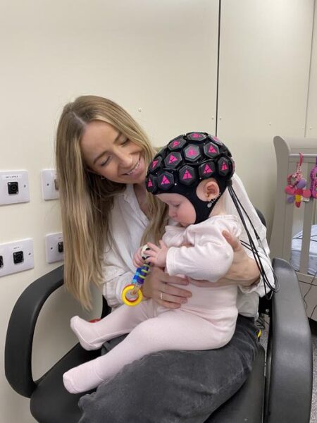
Summary: Researchers have developed a whole-head wearable brain imaging device that can map neural activity across an infant’s entire cortex, offering unprecedented insights into early brain development.
Estimated reading time: 8 minutes
A groundbreaking study led by researchers at University College London (UCL) and Birkbeck has unveiled a new wearable brain imaging technology that provides the most comprehensive view to date of infant brain activity in real-world situations. This innovative device, which uses harmless light waves to measure neural activity, has the potential to revolutionize our understanding of early brain development and neurodevelopmental conditions.
The study, published in Imaging Neuroscience, describes a significant advancement in high-density diffuse optical tomography (HD-DOT) technology. This new wearable headgear can measure brain activity across the entire outer surface of a baby’s brain, a vast improvement over previous versions that could only sample specific areas.
Shedding Light on Infant Brain Function
Dr. Liam Collins-Jones, the study’s first author from UCL Medical Physics & Biomedical Engineering and the University of Cambridge, explains the significance of this development: “The new method allows us to observe what’s happening across the whole outer brain surface underlying the scalp, which is a big step forward. It opens up possibilities to spot interactions between different areas and detect activity in areas that we might not have known to look at previously.”
This technological leap forward has already yielded surprising results. The researchers observed unexpected activity in the prefrontal cortex – an area associated with processing emotions – in response to social stimuli. This finding suggests that babies as young as five months old may already be processing social situations in complex ways.
Professor Emily Jones from Birkbeck, University of London, emphasizes the unique capabilities of this new technology: “This is the first time that differences in activity across such a wide area of the brain have been measured in babies using a wearable device, including parts of the brain involved in processing sound, vision and emotions.”
Real-World Applications and Future Potential
The researchers tested the device on 16 babies aged five to seven months. The infants watched videos of actors singing nursery rhymes and moving toys while sitting on their parent’s lap. This setup allowed the team to compare brain activity during social and non-social scenarios in a natural environment.
Why It Matters
This technology could have far-reaching implications for understanding typical and atypical neurodevelopment. By mapping brain activity and connections in infants, researchers may gain crucial insights into conditions such as autism, dyslexia, and ADHD at much earlier stages than previously possible.
Dr. Collins-Jones highlights the potential impact: “This more complete picture of brain activity could enhance our understanding of how the baby brain functions as it interacts with the surrounding world, which could help us optimise support for neurodiverse children early in life.”
Overcoming Limitations of Traditional Brain Imaging
Traditional brain imaging methods like magnetic resonance imaging (MRI) have significant limitations when it comes to studying infant brain activity. MRI requires subjects to remain very still, which is challenging for babies and limits the types of scenarios that can be studied.
The new HD-DOT technology offers several advantages:
- Portability: The device is wearable and doesn’t require a large, stationary scanner.
- Natural environments: Babies can be studied while interacting with parents or engaging in typical activities.
- Cost-effectiveness: The technology is less expensive than MRI, potentially making it more accessible for research and clinical use.
- Safety: The device uses harmless light waves instead of magnetic fields or radiation.
From Lab to Real World: Collaborative Innovation
The development of this technology showcases the power of collaboration between academic research and commercial development. The device used in the study was adapted from a commercial system developed by Gowerlabs, a UCL spin-out company founded by researchers from UCL’s Biomedical Optics Research Laboratory.
Dr. Rob Cooper, senior author of the study from UCL Medical Physics & Biomedical Engineering, notes: “This device is a great example of academic research and commercial technological development working hand-in-hand. The long-standing collaboration between UCL and Gowerlabs, along with our academic partners, has been fundamental to the development of wearable HD-DOT technology.”
As researchers continue to refine and apply this technology, it promises to open new avenues for understanding the intricate workings of the developing brain. This could lead to earlier interventions for neurodevelopmental conditions and more tailored support for all children as they grow and learn.
Quiz:
- What is the name of the brain imaging technology used in this study?
- At what age were the babies in the study?
- What unexpected brain activity did the researchers observe in infants?
Answer Key:
- High-density diffuse optical tomography (HD-DOT)
- Five to seven months old
- Activity in the prefrontal cortex in response to social stimuli
Further Reading
For more information on HD-DOT and its applications:
- HD-DOT Technology: https://www.ncbi.nlm.nih.gov/pmc/articles/PMC8033536/
- Optical Neuroimaging: https://en.wikipedia.org/wiki/Neuroimaging
To learn more about brain development in infants:
- Infant Brain Development: https://www.ncbi.nlm.nih.gov/pmc/articles/PMC3722610/
- Neurodevelopmental Disorders: https://www.chop.edu/services/neurodevelopmental-disorders-consultation-clinic
To explore the potential applications of this technology in clinical settings:
- Early Intervention for Neurodevelopmental Disorders: https://www.cdc.gov/autism/index.html
For additional news and research on wearable technology:
- Wearable Technology News: https://www.cnet.com/products/wearable-tech/
If our reporting has informed or inspired you, please consider making a donation. Every contribution, no matter the size, empowers us to continue delivering accurate, engaging, and trustworthy science and medical news. Independent journalism requires time, effort, and resources—your support ensures we can keep uncovering the stories that matter most to you.
Join us in making knowledge accessible and impactful. Thank you for standing with us!

