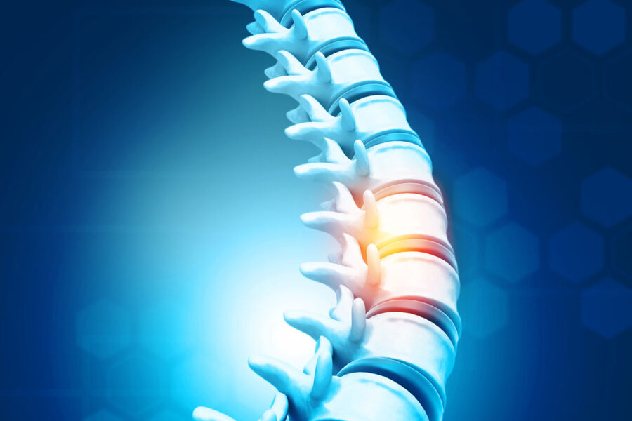
Summary: UCSF researchers have uncovered a molecular pathway that controls scar tissue formation after spinal cord injuries, potentially paving the way for future targeted drug treatments to promote optimal healing.
Estimated reading time: 6 minutes
UC San Francisco scientists have made a breakthrough in understanding how scar tissue forms after spinal cord injuries. Their research, published in Nature on September 18, reveals a previously unknown role of cerebrospinal fluid (CSF)-contacting neurons in controlling the balance of protective scarring.
Spinal cord injuries can have devastating consequences, often resulting in permanent nerve damage, loss of sensation, or paralysis. While initial scar formation helps protect and stabilize the injured area, excessive scarring can impede nerve regeneration and hinder recovery.
The Double-Edged Sword of Scar Tissue
After a spinal cord injury, nearby cells quickly form protective scar tissue around the damaged area. This immediate response is crucial for stabilization. However, over time, too much scarring can prevent nerves from regenerating, leading to permanent damage.
Current treatments for spinal cord injuries focus on stabilizing the spine through surgery or braces, managing pain and swelling with drugs, and physical therapy. However, these approaches do not directly address the complex biology of scar formation.
Unveiling the Role of CSF-Contacting Neurons
The UCSF team, led by David Julius, PhD, 2021 Nobel laureate in Physiology or Medicine, focused their research on a poorly understood group of neurons called cerebrospinal fluid (CSF)-contacting neurons. These cells line the central channel of the spinal cord and extend into the spinal fluid.
Using a novel method to label, isolate, and analyze these neurons, the researchers discovered that they express a receptor sensitive to κ-opioids, naturally produced by the human body. Further investigation revealed that κ-opioid signaling decreases after a spinal cord injury, triggering the transformation of nearby cells into scar tissue.
“By illuminating the basic signaling biology behind spinal cord scarring, these findings raise the possibility of one day being able to pharmacologically fine-tune the extent of that scarring,” said David Julius, senior author of the study and professor and chair of physiology at UCSF.
Balancing Act: Modulating Scar Formation
The research team’s experiments with mice yielded intriguing results. When they delivered extra κ-opioids to injured mice, scar formation decreased. However, this reduction in scarring came with a trade-off: the spinal cord injuries were more severe, and the mice showed poorer recovery of motor coordination.
These findings highlight the delicate balance required in the healing process. As Wendy Yue, PhD, first author of the paper and assistant professor of physiology at UCSF, explains: “κ-opioids might give us a way, after a spinal cord injury, to pharmacologically modulate the fine balance between producing enough scar tissue and having excessive scarring.”
Implications and Future Directions
This discovery opens up new possibilities for targeted treatments in spinal cord injuries. Unlike commercial opioid drugs such as oxycodone and hydrocodone, κ-opioids are generally not addictive, making them potentially safer for long-term use.
However, several questions remain to be answered before this research can be translated into clinical applications:
- Why do κ-opioid levels drop after spinal cord injuries?
- What are the ideal levels of scarring to support optimal healing?
- How can κ-opioid-related drugs be safely and effectively administered to humans with spinal cord injuries?
Further preclinical studies will be necessary to address these questions and pave the way for potential human trials.
The Value of Basic Scientific Research
This study underscores the importance of fundamental scientific inquiry. As Julius notes, “We were not looking for a way to control spinal cord healing. This came out of asking questions about this mysterious cell type, and then running into a mechanism that is both biologically interesting and could eventually have some therapeutic potential.”
By pursuing curiosity-driven research into poorly understood cellular mechanisms, scientists can uncover unexpected insights with far-reaching implications for human health and medicine.
Quiz
- What type of neurons did the UCSF researchers focus on in this study?
- What receptor did these neurons express that is relevant to scar formation?
- What happened when extra κ-opioids were delivered to injured mice?
Answers:
- Cerebrospinal fluid (CSF)-contacting neurons
- A receptor that senses κ-opioids
- Scar formation decreased, but spinal cord injuries were more severe and mice showed poorer recovery of motor coordination
Further Reading:
- Spinal Cord Injury – Mayo Clinic
- Opioid Receptors – StatPearls
- Neuroplasticity after Spinal Cord Injury
Glossary of Terms:
- Cerebrospinal fluid (CSF): Clear, colorless fluid that surrounds the brain and spinal cord, providing protection and nutrients.
- κ-opioids: Naturally produced molecules in the body that interact with specific receptors, distinct from commercial opioid drugs.
- Neuroregeneration: The regrowth or repair of nervous tissues, cells, or cell products.
- Scar tissue: Fibrous tissue that replaces normal tissue after injury, characterized by its lack of specialized structures.
- Spinal cord: The bundle of nerve fibers that runs down the middle of the back, carrying messages between the brain and the rest of the body.
- Preclinical studies: Research conducted using cell cultures or animal models before moving to human trials.
Enjoy this story? Get our newsletter! https://scienceblog.substack.com/
If our reporting has informed or inspired you, please consider making a donation. Every contribution, no matter the size, empowers us to continue delivering accurate, engaging, and trustworthy science and medical news. Independent journalism requires time, effort, and resources—your support ensures we can keep uncovering the stories that matter most to you.
Join us in making knowledge accessible and impactful. Thank you for standing with us!

