Scientists believed they understood what happens to DNA during cell division. The genome, they thought, sheds its intricate 3D architecture entirely, then slowly rebuilds it after the split. Now researchers at MIT have discovered this tidy picture was wrong.
Using a souped-up mapping technique that captures details 100 to 1,000 times finer than standard methods, the team found tiny loops persisting throughout cell division. These structures, dubbed microcompartments, actually grow stronger as chromosomes compact for division, then fade as cells return to normal life.
“We went into this study thinking, well, the one thing we know for sure is that there’s no regulatory structure in mitosis, and then we accidentally found structure in mitosis.”
Anders Sejr Hansen, an associate professor of biological engineering at MIT, admits the discovery caught his team off guard. They had set out to understand how microcompartments form by studying the moment they expected these structures to vanish: during mitosis, when cells divide.
When Packing Tight Creates Unexpected Connections
The researchers tracked mouse cells through the entire division process using a technique called Region-Capture Micro-C. At each stage, from initial division through the phase when daughter cells stabilize, they mapped which bits of DNA stuck together in three dimensions.
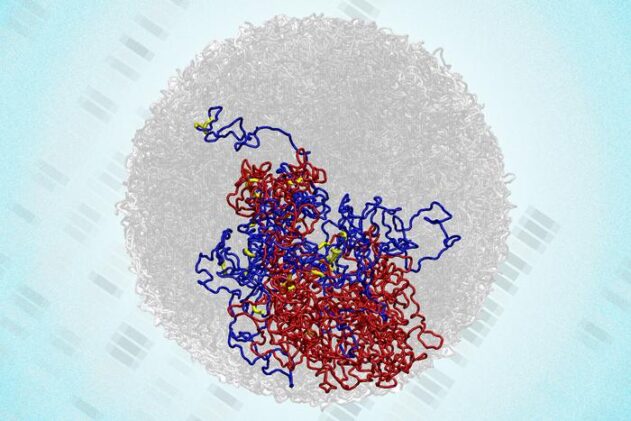
The big structures everyone knew about did disappear as expected. Compartments spanning millions of DNA letters, organizational domains, and large-scale loops all dissolved during division. But zooming in revealed something nobody had seen before: enhancers and promoters, the genetic switches that control when genes turn on, were forming tight little clusters.
These microcompartments peak in strength during anaphase and telophase, the phases when chromosomes become most compact. The finding makes physical sense. When you squeeze DNA into a smaller space, regulatory elements that normally sit far apart get pushed close together. If they have an affinity for sticking to one another, compaction gives them no choice but to connect.
Viraat Goel, lead author of the study published in Nature Structural and Molecular Biology, notes the work bridges a longstanding gap. Understanding how genome structure translates to function, specifically how genes get switched on and off, has puzzled researchers for decades.
An Accidental Burst of Activity
The discovery might explain a phenomenon that has mystified cell biologists since the 1960s. Back then, everyone assumed gene transcription shut down completely during division. But studies from 2016 and 2017 showed something odd: many genes experience a brief spike of activity near the end of mitosis before being suppressed again.
The MIT team found that genes showing this transcriptional spike were more likely to sit at microcompartment anchors. The loops appear to activate transcription somewhat by accident, creating favorable conditions for gene expression before the cell quickly shuts things down.
“It almost seems like this transcriptional spiking in mitosis is an undesirable accident that arises from generating a uniquely favorable environment for microcompartments to form during mitosis.”
Once division completes and cells enter a resting phase called G1, many of these small loops weaken or disappear. The researchers suspect cells actively prune unwanted connections, keeping only the regulatory loops needed for proper gene expression.
Computer simulations helped explain what drives microcompartment behavior. Three factors matter: how strongly regulatory elements attract each other, how densely packed the chromosomes are, and how much other molecular machinery interferes by creating competing structures. During the peak of division, compaction is high and interference is low, creating ideal conditions for microcompartments.
The finding suggests cells might use chromosome compaction as a global dial to tune gene regulation, not just as a way to organize DNA for division. Hansen and colleagues are now exploring whether variations in cell size and shape, which affect how tightly DNA packs, might explain some previously mysterious changes in genome organization.
The research was funded by the National Institutes of Health, a National Science Foundation CAREER Award, the Broad Institute, a Pew-Steward Scholar Award, the Mathers Foundation, the MIT Westaway Fund, the Koch Institute Bridge Project, and the Koch Institute Support Grant from the National Cancer Institute.
Nature Structural & Molecular Biology: 10.1038/s41594-025-01687-2
ScienceBlog.com has no paywalls, no sponsored content, and no agenda beyond getting the science right. Every story here is written to inform, not to impress an advertiser or push a point of view.
Good science journalism takes time — reading the papers, checking the claims, finding researchers who can put findings in context. We do that work because we think it matters.
If you find this site useful, consider supporting it with a donation. Even a few dollars a month helps keep the coverage independent and free for everyone.